
The ability to assess the performance of novel, non-toxic coatings is essential to developing environmentally benign solutions to control biofouling, which is the accumulation of organisms on man-made structures in aquatic environments. In the past decade, the methods for screening experimental coatings have increased and subsequently improved the ability to down-select the best candidate coatings rapidly. The standard approaches involve a series of assays that first examine the efficacy of non-toxic coatings as antifouling surfaces by quantifying the settlement of algal spores and invertebrate larval stages. Following settlement assays, coatings are often tested for their efficacy at limiting adhesion (i.e. ‘foul-release’ performance) by using a variety of techniques and marine organisms, including bacteria, algae and invertebrate larval and adult stages. This overview serves to describe the current techniques and technologies used in assessing marine coatings and acts as a gateway into the primary literature.
1 Introduction
Biofouling, the accumulation of organisms on submerged structures, continues to be a problem of significant economic and environmental concern for marine-dependent industries.1–4 Despite the ubiquitous nature of the problem, effective solutions to control biofouling over long periods of time are primarily limited to the use of antifouling measures, principally paints that contain heavy metals or other biocides, which are toxic to marine organisms and effectively limit their attachment and subsequent growth. Regulations preventing the use of toxic paints (e.g. paints containing tributyltin) and regulations limiting the concentrations of heavy metals in bays and harbors have spurred the development of environment-friendly fouling-resistant marine coatings (see Callow and Callow5 for a recent review of the development of non-toxic coating technologies). The focus of this overview is to provide a general description of the various laboratory and field methods used by the research community to evaluate and then down-select the best-performing coatings. The first review of this topic6 treated only laboratory approaches and focused on two organisms used in screening, the barnacle Amphibalanus (=Balanus) amphitrite and the green alga Ulva linza. A much more extensive and detailed overview of laboratory assays was provided by Briand7 in 2009, and a comprehensive and detailed edited volume was published by Dobrestov et al.1 in 2014. A major development in the field about a decade ago was the advent of combinatorial approaches to creating coatings,8 which necessitated an expansion of the types of assays used to evaluate novel formulations and the rapidity with which coatings could screened. For detailed information about individual assays or methods including, for example, concentration of test organisms and duration of assays, one should refer to the specific references mentioned in each section. The intent here is to provide an overview of evaluation methods and how each method contributes to the development of new coatings and the fundamental understanding of how non-toxic coatings function. Before introducing the various laboratory assays and field tests, a brief description of the life cycles of biofouling organisms will be provided.
1.1 General description of life cycles of biofouling organisms
Most marine fouling organisms, including, for example, protozoans, algae and invertebrates, possess a life cycle with multiple stages – that is, biofouling organisms possess what is termed a ‘complex life cycle’. In this type of life cycle, there is usually a microscopic free-swimming life stage (a larva in the case of many invertebrates, or a spore in the case of many algae). The free-swimming stage must transition from swimming in the water to living on a surface such as rock and sand or a man-made structure such as the hull of a ship. The initial transition from the free-swimming phase to the attached adult phase is a process referred to as settlement. The organism at the time of settlement will attach to a surface by using an adhesive. It will then undergo development into the next stage of the life cycle, typically the sexually reproductive adult stage. Bacteria and other microorganisms, although without a complex life cycle, attach to submerged objects as well. Collectively, the accumulation of the organisms on a man-made surface is biofouling. What follow from a biological understanding of these processes are two strategies for intervening and changing the process of biofouling: (a) the presettlement process, which prevents the initial settlement and attachment of organisms (i.e. antifouling strategy), and (b) the postsettlement process, which disrupts the adhesion of organisms after settlement (i.e. foul-release strategy). A non-toxic coating can work at one or both of these stages in biofouling development, and indeed, the ideal coating likely prevents both the settlement and permanent attachment of biofouling organisms.
1.2 Organisms commonly used to screen marine coatings
There are a variety of organisms covering a large range of sizes and taxonomic groups commonly used in screening non-toxic coatings. Various species of bacteria are used to test adhesion properties; two species of algae, the spores and young plants of the green alga Ulva and the diatom Navicula, are used to assess antifouling and foul-release properties, as are three invertebrate species, the adults and larvae of the tube worm Hydroides elegans, the adults and larvae of the barnacle A. amphitrite and adult mussels (Mytilus sp.) (Figure 1).5
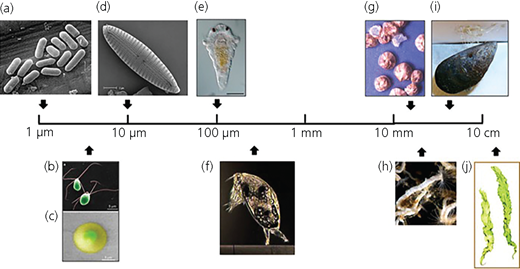
Typical species used to test coatings showing diversity and size scales: (a) bacteria (scanning electron micrograph (SEM)), (b) false-color SEM of motile, quadriflagellate spores of the green alga (seaweed) Ulva, (c) false-color environmental SEM image of settled spore of Ulva showing secreted annulus of swollen adhesive, (d) SEM of diatom (Navicula), (e) larva of tube worm, H. elegans (image courtesy of B. Nedved), (f) barnacle cypris larva (A. amphitrite) exploring a surface by its paired antennules (image courtesy of N. Aldred), (g) adult barnacles (image courtesy of A. S. Clare), (h) adult tube worms (H. elegans; image courtesy of M. Hadfield), (i) adult mussels showing byssus threads attached to a surface (image courtesy of J. Wilker) and (j) individual plants of the green alga (seaweed) Ulva. The diagram is intended to indicate relative scales rather than absolute sizes; individual species within a group can vary significantly in absolute size. From Callow and Callow5
Typical species used to test coatings showing diversity and size scales: (a) bacteria (scanning electron micrograph (SEM)), (b) false-color SEM of motile, quadriflagellate spores of the green alga (seaweed) Ulva, (c) false-color environmental SEM image of settled spore of Ulva showing secreted annulus of swollen adhesive, (d) SEM of diatom (Navicula), (e) larva of tube worm, H. elegans (image courtesy of B. Nedved), (f) barnacle cypris larva (A. amphitrite) exploring a surface by its paired antennules (image courtesy of N. Aldred), (g) adult barnacles (image courtesy of A. S. Clare), (h) adult tube worms (H. elegans; image courtesy of M. Hadfield), (i) adult mussels showing byssus threads attached to a surface (image courtesy of J. Wilker) and (j) individual plants of the green alga (seaweed) Ulva. The diagram is intended to indicate relative scales rather than absolute sizes; individual species within a group can vary significantly in absolute size. From Callow and Callow5
2 Screening assays of settlement inhibition (i.e. antifouling)
The most common method by which coatings are evaluated for their efficacy at inhibiting settlement is through various laboratory-based ‘settlement assays’ using algae and invertebrates. The standard substratum for screening is a coated glass slide. Coatings should be of uniform thickness and well bonded to the glass slides. Multiple replicates are used (generally three to six) to ensure that observed differences among coatings can be statistically validated.
In the case of green algae, adult U. linza plants are collected from the shore and immersed in seawater, after which motile spores are released. The spores are on the order of 5–8 μm and will adhere themselves to a suitable surface.6 The spores demonstrate ‘preferences’, which leads to different settlement rates depending on the topographical9–11 or physical/chemical12–15 properties of the surface. To run assays, slides are placed in individual compartments of quadriPerm dishes to which zoospore suspension is added. The dishes are incubated to allow settlement and attachment. After the incubation period, unattached spores are removed and the attached spores are enumerated using microscopy. Comparative data are generally reported as density of spores per square millimeter for each tested coating (Figure 2).16 Detailed protocols and settlement studies can be found in the early work by Callow et al.17
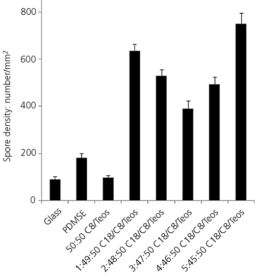
Settlement of zoospores of Ulva on xerogel coatings and glass and polydimethylsiloxane elastomer (PDMSE) standards. Each value is the mean of 90 counts on each of three replicate slides. Error bars represent 95% confidence limits. Teos, tetraethoxysilane. From Gunari et al.16
Settlement of zoospores of Ulva on xerogel coatings and glass and polydimethylsiloxane elastomer (PDMSE) standards. Each value is the mean of 90 counts on each of three replicate slides. Error bars represent 95% confidence limits. Teos, tetraethoxysilane. From Gunari et al.16
Other common settlement assays often utilize the purple-striped barnacle A. amphitrite.6,18 Rittschof et al.18 produced the foundational knowledge for the field by developing the mass laboratory culturing techniques used to produce the terminal stage cypris larva of A. amphitrite (Figure 1) and by developing toxicity and settlement inhibition assays for larvae. Currently the most common procedure uses small volumes (c. 0·5 ml) of seawater containing 20–50 larvae that are placed directly on the surface of a coating in what is termed a ‘droplet assay’. The slides are typically in a covered container (e.g. a petri dish) to prevent evaporation and placed in a temperature- and light-controlled incubator for 24–48 h, after which the number of swimming larvae and attached barnacles are enumerated. The data are usually displayed as a percentage of individuals that settled in each condition. Control treatments (e.g. plain glass slides or polydimethylsiloxane elastomer (PDMSE)) are generally run in parallel with the experimental coatings to ensure that the larvae are competent to metamorphose (Figure 3).16 The droplet assay is the most commonly used settlement assay for barnacles. In certain circumstances, such as when a coating is extremely hydrophobic or hydrophilic, coated petri dishes or vials are used in place of glass slides.19 The duration of experiments and analysis and presentation of data are similar to the droplet assay.
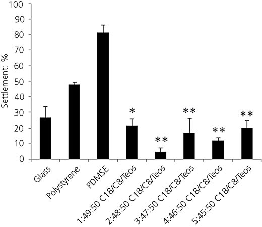
Settlement of barnacle cyprids on xerogel coatings applied to glass dishes, polystyrene and glass standards. Each value is the mean from three replicate measurements. Error bars represent the standard error (SE) of the mean. *, values which are significantly lower than the polystyrene standard; **, values which are significantly lower than the glass and polystyrene standards. From Gunari et al.16
Settlement of barnacle cyprids on xerogel coatings applied to glass dishes, polystyrene and glass standards. Each value is the mean from three replicate measurements. Error bars represent the standard error (SE) of the mean. *, values which are significantly lower than the polystyrene standard; **, values which are significantly lower than the glass and polystyrene standards. From Gunari et al.16
2.1 Understanding antifouling mechanisms
An inherent limitation of settlement assays is that one does not often learn the mechanism by which the coatings are working. That is, what factors mediate the antifouling characteristics of non-toxic coatings? Recent developments in equipment and software have enabled tracking of larval and spore behavior, thus addressing this important shortcoming of traditional settlement assays. For example, Heydt et al. 20 developed four-dimensional holographic tracking for algal spores. Their work enables quantification of various motility patterns and the frequency of different patterns in response to different coating surface chemistries. The technique has been utilized to understand the settlement response of U. linza zoospores to cationic oligopeptide surfaces (Figure 4).21 Invertebrate larvae are significantly larger than algal spores (c. 200 μm against 10 μm, respectively) which presents challenges in trying to quantify behaviors that occur in a relatively large field of view. Initial work was done using two-dimensional (2D) tracking software, EthoVision 3.0, whereby larval settlement behavior was quantified for individual larvae that were in contact with or close to a surface.22 The affinity of larvae for different chemistries could be quantified by the amount of time the larvae spent exploring a surface and the pattern of their movements.23 More recent research has extended the 2D analysis to three dimensions by using stereoscopy.24 This technique has allowed quantification of swimming behavior at various distances away from a surface, which can be correlated with the settlement preferences of larvae for surfaces with different chemistries (Figure 5).24 The technique of tracking the behavior of larvae and spores has enabled a better understanding of how novel, non-toxic coatings function and has provided insight into the fundamental understanding of organismal settlement behavior.
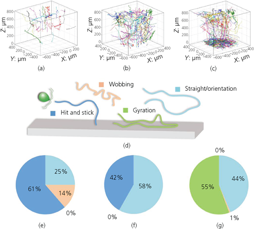
Three-dimensional (3D) spore trajectories and pattern occurrence on surfaces with different ArgTyr concentrations. Spore trajectories are shown as 3D representations for (a) 100, (b) 50 and (c) 0% ArgTyr surfaces. (d) Schematic representation of the different motion patterns. (e–g) Occurrence of the motion patterns ‘hit and stick’, ‘wobbling’, ‘gyration’ and ‘straight path’/’orientation’ for the analyzed trajectories above the respective surfaces within an observation time of 2 min. From Vater et al. 21
Three-dimensional (3D) spore trajectories and pattern occurrence on surfaces with different ArgTyr concentrations. Spore trajectories are shown as 3D representations for (a) 100, (b) 50 and (c) 0% ArgTyr surfaces. (d) Schematic representation of the different motion patterns. (e–g) Occurrence of the motion patterns ‘hit and stick’, ‘wobbling’, ‘gyration’ and ‘straight path’/’orientation’ for the analyzed trajectories above the respective surfaces within an observation time of 2 min. From Vater et al. 21
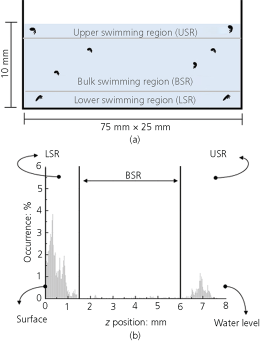
(a) Schematic side-view of the test container and (b) a representative histogram of the z position of active (velocity v > 0 mm/s) cyprids showing the main swimming regions. From Maleschlijski et al. 24
(a) Schematic side-view of the test container and (b) a representative histogram of the z position of active (velocity v > 0 mm/s) cyprids showing the main swimming regions. From Maleschlijski et al. 24
3 Screening assays to assess foul-release properties of coatings
The initial set of screening assays on coatings focuses on understanding their antifouling properties. Although it would be ideal to have complete efficacy at the time of settlement of spores and larvae, it remains clear that organisms will accumulate and that non-toxic coatings must function as ‘foul-release coatings’ – chemistries that disrupt the natural adhesion process such that organisms are released under hydrodynamic stress created as water flows over the surface of a coating. A variety of laboratory and field assays have been developed to measure and compare the relative adhesion of organisms to marine coatings. What follows is an overview of the various approaches currently utilized.
3.1 Assays of bacterial adhesion
The quantification of bacterial fouling to test the performance of coatings has been done for many decades.13,25–30 In the development of marine coatings, there are a variety of approaches used, which most commonly involve (a) incubating a monoculture of bacteria with a coated surface to allow cell attachment, (b) enumeration of the attached cells, (c) application of a known shear force by repeated dipping of a slide in water,29 placing biofilmed surfaces in a flow cell13,31 or attaching biofilmed surfaces to a rotating drum29,32 and then (d) enumeration of the remaining attached bacteria. The data are often presented as the quantity of cells or biofilm initially attached and then the per cent removal after application of a shear force (Figure 6).29
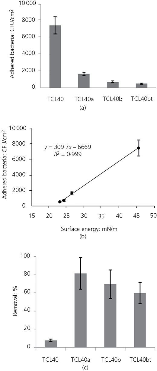
(a, b) Formation and (c) removal of Pseudomonas fluorescens biofilm on silicon oxide (SiOx) coatings. The correlation between bacterial attachment and surface energy is shown in (b). N = 5; error bars are 2 × SE. CFU, colony-forming units. From Akesso et al. 29 TCL40, TCL40a, TCL40b and TCL40bt are coatings with decreasing O2:hexamethylsiloxane ratios
(a, b) Formation and (c) removal of Pseudomonas fluorescens biofilm on silicon oxide (SiOx) coatings. The correlation between bacterial attachment and surface energy is shown in (b). N = 5; error bars are 2 × SE. CFU, colony-forming units. From Akesso et al. 29 TCL40, TCL40a, TCL40b and TCL40bt are coatings with decreasing O2:hexamethylsiloxane ratios
Another recently developed high-throughput assay involves measuring the retention and then retraction of a developed biofilm on a coating surface.28 The technique involves incubating coated multiwell plates with bacterial suspensions to allow biofilms to develop. Following incubation, the biofilms retained on the coating surface after washing are stained with crystal violet (CV). The stained biofilm is then allowed to dry. During the drying process, the biofilm, depending on the foul-release efficacy of the coating, will retract to a smaller surface area (Figure 7).28 The degree of retraction from the hydrated biofilm is measured as a per cent coverage; no difference between the area of the hydrated and dried biofilm would be calculated as 100% coverage. Importantly, the amount of retraction has been shown to be correlated directly with barnacle adhesion data gathered from co-occurring field experiments.33
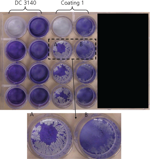
Image of CV-stained polysiloxane coating 1 (A) after drying and (B) before drying. Similarly, the left column of Dow Corning (DC) 3140 was CV stained after drying; the right column, before drying. From Stafslien et al. 28
Image of CV-stained polysiloxane coating 1 (A) after drying and (B) before drying. Similarly, the left column of Dow Corning (DC) 3140 was CV stained after drying; the right column, before drying. From Stafslien et al. 28
3.2 Assays of algae and invertebrate adhesion
3.2.1 Water jet
Because of the relative sizes of algae and invertebrates (Figure 1), a different series of techniques is often used with such organisms to assess the foul-release efficacy of coatings. The primary techniques utilized are water jet, in both the laboratory and field,34–38 flow cells39,40 and handheld and automated force gauge for removal of animals in shear.41–44
An early published account using a water-jet test came from Rittschof et al.34 as part of their study to develop rapid field assessment of coatings. The technique involved exposing coated fiberglass rods (110 mm × 7 mm) to natural fouling communities. After fouling occurred, the rods were weighed, sprayed with a calibrated Waterpik and weighed again to determine how much fouling was removed. In this case, the rods are exposed in the field and then fouling is removed under laboratory conditions. An entirely field-based water-jet system was originally developed by Swain and Schultz35 as a mechanism to test the adhesion of biofilms and soft fouling that developed during exposure of coatings at static field sites. Briefly, the apparatus consists of a scuba tank containing compressed air connected to a regulator with a valve used to vary pressure (typically 0–1·65 MPa). The regulated line pressurizes another scuba tank filled with seawater, off of which a blowgun is operated to create the water jet. The operator increases the pressure in a stepwise fashion and records at what pressures organisms are removed. This valuable tool has been utilized for almost two decades to evaluate the performance of marine coatings under field conditions. Several laboratory-based systems have been further developed for removing algal spores and young plants of U. linza,37,45,46 the diatom Navicula perminuta37 and barnacle cypris larvae of A. amphitrite.38 Cassé et al.37 developed as part of a high-throughput method a water-jet system using a 24-well plate format for removal of algal spores (U. linza) and a diatom (N. perminuta) and a method using coated microscope slides to assess adhesion of young plants of Ulva (also known as sporelings). The data generated from water-jet analyses are similar regardless of the particular technique utilized and generally involve showing the per cent removal as a function of impact pressure (Figure 8).37 To a lesser extent the pressure at which fouling is removed is reported and this is primarily done for data collected under field conditions.35
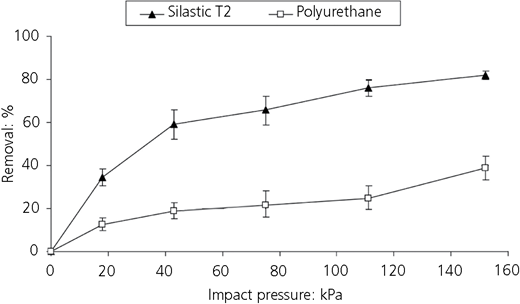
Percentage removal of Ulva sporeling after 5 d growth on coatings deposited in 24-well plates and hosed at 18, 43, 75, 111 and 152 kPa impact pressure with the Spinjet. Each point is the mean of six replicate wells. Bars represent ±95% confidence limits derived from arcsine-transformed data. From Cassé et al.37
Percentage removal of Ulva sporeling after 5 d growth on coatings deposited in 24-well plates and hosed at 18, 43, 75, 111 and 152 kPa impact pressure with the Spinjet. Each point is the mean of six replicate wells. Bars represent ±95% confidence limits derived from arcsine-transformed data. From Cassé et al.37
3.2.2 Flow channel
Another method used for hydrodynamic removal of algae and invertebrates is the use of a flow channel that develops a fully turbulent flow, generating a wall shear stress that removes organisms from the surfaces of coated slides.6,36,39 Spores and young plants of the green alga U. linza are commonly used with a flow-channel apparatus, as is the diatom N. perminuta.19 As with the water jet, cell, spore or sporeling density or biomass is quantified initially. The slides are then placed in the flow-channel apparatus and wall shear stresses are generated through turbulent flow that is created along a low-aspect ratio section of the channel just preceding the experimental slides.36,39,47 Following exposure to the turbulent flow, biomass is again quantified and the data are displayed as a per cent removal from the surface. Glass controls are commonly used, which show virtually 0% removal, as are standardized foul-release coatings, which show about a 50% reduction in biomass after being exposed to turbulent flow.48 Although not used routinely for invertebrate larvae and young juveniles, the flow channel has been used to examine adhesion of four species.40 Larvae of a barnacle (A. amphitrite), an ascidian (Phallusia nigra), a bryozoan (Bugula neritina) and a polychaete (H. elegans) were allowed to attach and metamorphose to surfaces. The individuals attached to each slide were enumerated and then exposed to turbulent flows for 4 min, after which the remaining individuals were enumerated. The data are presented as a percentage remaining attached.
3.2.3 Force gauge
Another common test of adhesion in screening of marine coatings is the removal of hard-fouling organisms by applying a shear force using a handheld (field) or automated (laboratory) force gauge. The method was first developed by Swain et al.41 for removal of barnacles in the field and has been designated as a standard test method.42 Since then, the method has been adapted to measure critical removal stress (originally termed ‘adhesion strength’) for other hard-fouling animals, including tube worms, encrusting bryozoans and oysters.49 The method has also been augmented and incorporated into laboratory-based assays.43,44,50
The original method developed to assess the foul-release properties of non-toxic coatings used a handheld probe to apply force to the base of a barnacle that is attached to a surface. The probe consists of a small stainless steel plate (14 mm × 14 mm as per ASTM D 5618-94 A42) which is connected to the end of an 8 mm dia. stainless-steel rod. The probe is attached to a commercial force-measuring device such as those produced by Imada, Inc. The basic procedure involves measuring the area of attachment of a fouling organism such as a barnacle, applying a shear force parallel to the base of the organism at a rate of 4·5 N/s and then recording the maximum force applied to remove the animal. The critical removal stress is then calculated by normalizing the maximum force during removal by the area of attachment.
The basic method developed for field applications has been modified by incorporation of automation first in the application of shear force43 and then later to become fully automated.44 The modification by Wendt et al.43 uses an automated test stand (Imada model SV-5) to deliver a constant rate of force and a reproducible angle of application; both items are critical to reducing variability among measurements of critical removal stress. In this case, the animals are also removed while continuously submerged in water. Data from the force gauge can be streamed using a digital interface, allowing creation of force against time curves used in investigating modes of release and fracture mechanics.43,51–53 The next stage in the development of the technique was automation of the entire system to facilitate rapid throughput of many coating formulations and less variability than the handheld method.44 In this computer-controlled technique, the instrument has a camera that is placed above a motorized platform on which an experimental coating is affixed. The camera takes an image of each barnacle on the coating from which the surface area of attachment is digitally quantified. The experimental surface is moved at a constant rate toward a fixed carbon fiber push bar that is 0·1 mm above the experimental surface. The push bar is connected to a calibrated load cell whose electrical output is converted to a force. Peak force and force against time curves are automatically captured for each barnacle, and the instrument’s software calculates critical removal stress.
3.2.4 Reattachment assays
A final method recently developed to assess the foul-release performance of non-toxic coatings involves removal and then reattachment of adult barnacles to coatings. The method was initially introduced by Rittschof et al.54 as a tool to study adhesion of barnacles to toxic coatings, which prevented normal settlement and growth. Being less sensitive than larvae and juveniles, adults could be reattached to coatings that would have otherwise been hard to test. The technique was later improved to enable a more rapid throughput of coatings since it only takes 1–2 weeks for a barnacle to reattach, whereas growth from a juvenile takes 1–2 months.55 Thus, coatings can be evaluated more rapidly than growing animals from juvenile stages. The basic method involves removing adults from a silicone elastomer (e.g. Dow Corning Silastic T2), drying the basal plate of the animals and then placing animals on a new experimental coating first in air for several hours to enable initial attachment, followed by immersion in filtered seawater and rearing according to standard husbandry methods.54 Within 7 d, the reattached animals require the same amount of force to remove from coatings compared to animals reared from juvenile stages.54
4 Correlation among laboratory and field assays
A key question centers around the ability of laboratory assays to predict performance in the field and, ultimately, in commercial applications. The simplicity and rapidity of the laboratory assays is both an asset and a liability in that they are relatively quick and easy, but they grossly simplify the dynamics of fouling in the marine environment. The complexity of fouling communities in the field is significant and results from dynamics of physical factors such as temperature and salinity, ecological factors such as the variability of larval and spore supply, and biological factors such as species interactions through predation and competition for area. It is within this complex system that marine coatings must perform and be tested. However, it is simply not practical in developing novel coatings, some of which may be very expensive or hard to produce in large quantities, to begin field-testing straight away. Moreover, the sheer number of formulations produced with the advent of combinatorial approaches necessitates initial screening in the laboratory. The most thorough approach to date to examining the relationship between the performances of coatings in the laboratory and field was conducted by Stafslien et al.33 In their study, eight novel siloxane–polyurethane coatings were assessed for their performance both in the laboratory and at three field sites: Morro Bay, California; Indian River Lagoon, Florida; and Tropical Marine Sciences Institute in Singapore. A suite of laboratory assays was performed at North Dakota State University, including adhesion assessments of bacteria, microalgae, macroalgal spores and adult barnacles. At the same time, coated panels were distributed for field evaluations utilizing standardized testing methods during a 6-month exposure. The results showed good agreement between the laboratory screening assays and the field assessments, with both laboratory and field-testing distinguishing the same best-performing coatings. The biofilm retraction assay and barnacle reattachment assays were shown to be the most predictive of broad-spectrum field performance. Thus, it is clear that laboratory assays are useful and informative in the down-select process.
5 Conclusions
Biofouling has major economic and environmental impacts. The ability to assess the performance of novel, non-toxic coatings is essential to developing environmentally benign solutions to control biofouling. In the past decade, the ability to screen coatings by using a variety of both laboratory and field techniques has grown vastly and subsequently improved the ability to down-select the best candidate coatings rapidly. Many methodologies for screening coatings in the laboratory have become automated, allowing for a high throughput of coating formulations. The standard methodologies involve a series of assays that first examine the efficacy of non-toxic coatings as antifouling surfaces by quantifying the settlement of algal spores (e.g. the green alga U. linza) and invertebrate larval stages (e.g. barnacle cyprids). The data from settlement assays determine how stimulatory or inhibitory the coatings are relative to known standards. Following the settlement assays, coatings are often tested for their efficacy at limiting adhesion (i.e. their foul-release properties). Foul-release assays involve a variety of organisms, including bacteria, algae and invertebrates, using several technical approaches, including water jet, calibrated flow cells and handheld or automated force gauges. Results from the foul-release assays determine how well the coatings limit adhesion and their potential for releasing fouling under hydrodynamically induced shear forces (e.g. flow created as a ship moves through the water). The robust suite of bioassays available in both the laboratory and field enables a richer understanding of the physical and chemical properties of coatings that lead to effective non-toxic strategies to control biofouling.
Acknowledgements
The author would like to acknowledge funding from the US Office of Naval Research (ONR) (award number N00014-12-1-0432) and the colleagues and collaborators that have participated in the ONR Biofouling Program over the past 15 years.
