


The topology of the action potential
The topology of the action potential
KeywordsCybernetics, Neurosciences
Abstract Discusses the topology of the action potential which is a wave of depolarisation which travels along the axon of a nerve. Describes the action of the ion channels in medullated nerves, and offers explanations as to their roles.
The action potential is a wave of depolarisation which travels along the axon of a nerve, and since a fast response to any stimulus is advantageous the action potential is presumably optimised in that direction. The number of cortical neurones in man is currently estimated as 28 billion, these are arranged in columns of six cells of which 60 per cent are pyramidal cells with long axons. The resting potential is about 0.08 volts but the thickness of the cell membrane is so small that the field is variously estimated at 10,000 to 100,000 volts per centimetre. This is a very high field indeed as the breakdown field of olive oil is 10,000 volts when the radius of the electrodes is about 2cms, the axons have a radius of about a micron so the integrity of the insulation is impressive. The resting potential is maintained by ion pumps using ATP as a fuel. If the insulation is broken down a wave of depolarisation travels in both directions along the axon. The depolarisation wave of action potential is propagated by the opening of ion channels in the cell membrane. The resting potential is thought to keep the contents of the cell from escaping and its loss usually signifies the death of the cell.
In medullated nerves the ion channels are concentrated at the nodes of Ranvier, which act as repeaters on the axon. The number of ions which pass through a channel is measured in thousands so they must form a mushroom cloud within the axon, and the total of the channels in a section across the axon at the point of breakdown will form a double torus, the major torus being downstream and the minor in the wake of the wave (Figure 1). The channels are distributed in a slightly random manner so that small irregularities in the shape of the major torus may occur; when these cause a forward projection propagation will be slowed by the weakening of the field at that point and, conversely, when there is a slight invagination the field will be strengthened so the wave will tend to keep the double toroidal shape.
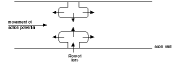
Figure 1.Flow of ions
The meduallation has a laminated structure suggestive of a superlattice. This is in effect an interference filter vertical to its surface but a wave guide along its axis so the action potential may even travel at the speed of light between one node and the next; this is difficult to determine since the delay at the node is unknown. Owing to the toroidal shape of the ion cloud within the axon there is a longitudinal component of the field along the axis of the axon. The channels are rings of polypeptides placed normal to the axon wall and embedded in the cell membrane. The polypeptide molecules are held in position mainly by weak hydrogen bonds and held vertical to the cell membrane by the resting potential, the longitudinal component of the torus field will cause the polypeptides to tilt, possibly causing them to become misaligned so that the ion channel is opened and the action potential can be propagated. The passage of ions through a channel constitutes a flow of particles through an electric field and since every particle has a wave and every wave a particle the opening of a channel will produce photons with an energy of 80mEv in the near IR of the light spectrum.
These would be strongly absorbed by water in the vicinity but there is no reason why with suitable technology they should not be observed. Since the tori are symmetrical, in theory they should cancel out when perfect electrodes are placed symmetrically on either side of the axon, but fortunately, perfect electrodes cannot be constructed so the signal can be detected. Individual channels produce small spikes but the toroidal shape of the action potential wave causes the recorded shape to become curved. The action potential is generated at the axon hillock, the constriction at the beginning of the axon. Its generation is determined by the integral of all the stimulatory and inhibitory potentials produced upstream in the dendritic field so the action potential may be considered as a vote taken by the conflicting views of the 4,000 constituents. The action potential is propagated downstream in the wall of the axon but also upstream in the lumen of the dentrites so that there is feedback to the input synapses which might alter their weighting, an embodiment of Hebb's law that the thresholds to stimulus fall with repeated use. The combined upstream dendritic field constitutes a decoder, the combined downstream field must constitute an encoder, for information to be encoded in the pattern of different synaptic weightings both input and output synapses must possess some element of plasticity which may be controlled by feedback, rather like a miniaturised white rat in a Skinner box,or, in electronic parlance, a Boltzmann machine.
Brennig JamesCherry Orchard, Marlow Common, Bucks SL7 2QP
The authors would like to thank Mr N. Barrow, Miss L. Lee, and other colleagues at Manchester Metropolitan University, for this paper would not have been finished so quickly without their warm and valuable help. This project is supported by Fok Ying Tung Education Foundation.
Editor's note: Communications and forum contributions are not sent to referees and consequently will be available to readers much more quickly. Comments and alternative viewpoints on all matters pertaining to cybernetics and systems are sought, particularly on the many issues that are raised in this section.

