Computed tomography (CT) is a significant clinical detection method without invasive procedures. However, high-dose radiation is associated with elevated risk of cancer. Low-dose CT reconstruction techniques have the potential to significantly reduce this risk. These techniques reduce radiation dose by two means: low radiation intensity and incomplete scans. The focus of this review is incomplete-scan CT reconstruction techniques, including sparse-view and limited-angle CT reconstruction. In the context of incomplete scans, CT images obtained by conventional CT reconstruction techniques exhibit noises and streak artifacts. Deep learning techniques are changing this situation. However, there are few reviews that recently focus on the fast developing area. This paper reviews recent research on CT reconstruction of incomplete scans via deep learning and classifies them into two categories including image domain and reconstruction domain processing methods. This study also evaluates the strengths and limitations of these approaches and discusses future prospects, highlighting promising technologies and directions.
1. Introduction
Computed tomography (CT) is an important noninvasive imaging technique used to examine the internal structures of the human body. It offers advantages such as rapid scanning speed and high image resolution, making it a vital tool in clinical medical diagnosis (Brenner and Hall, 2007; Katsuragawa and Doi, 2007). Most CT systems utilize X-rays as the imaging source due to their ability to penetrate solid materials (Kak and Slaney, 2001). During a CT scan, an X-ray source captures multiple two-dimensional (2D) projections from different angles, measuring the attenuation of X-rays through tissues to produce high-quality cross-sectional images that assist physicians in accurate diagnosis. However, prolonged exposure to high doses of X-ray radiation may lead to metabolic abnormalities and even increase the risk of cancer (Hall and Brenner, 2008). Therefore, reducing the radiation dose while maintaining reconstructed image quality has become a major research focus.
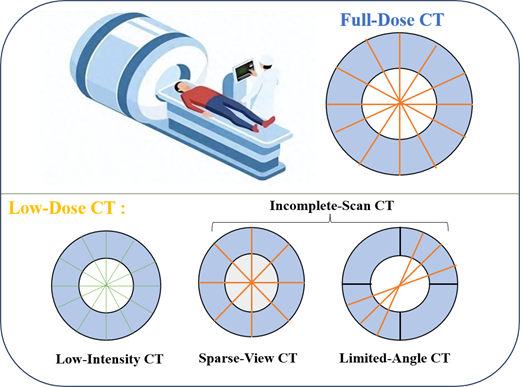
Current approaches to reducing radiation dose in CT scans (Kulathilake et al., 2023), as shown in Figure 1, can be broadly categorized into two types. The first approach involves lowering the tube voltage and current to reduce radiation intensity, which is referred to as low-dose CT reconstruction. The second approach maintains the radiation intensity but decreases the number of projection angles (sparse-view CT reconstruction) or restricts the angular range of scanning (limited-angle CT reconstruction). This review primarily focuses on the second category. It is crucial to note that while limited-angle and sparse-view CT are both challenged by incomplete data, their nature differs fundamentally. Limited-angle CT suffers from a complete absence of projections within a angular range, leading to severe global distortions. Whereas sparse-view CT, with its insufficient angular sampling, is primarily plagued by streak artifacts. Consequently, the optimal priors and reconstruction strategies are distinct and must be chosen accordingly.
Conventional CT reconstruction methods include the filtered back projection (FBP) algorithm (Shepp and Logan, 1974) and iterative reconstruction (IR) algorithms (Andersen and Kak, 1984). However, when dealing with undersampled data from sparse-view or limited-angle acquisitions, these classical approaches often introduce severe artifacts and noise into the reconstructed images, which are unacceptable in clinical practice.
In recent years, deep learning technology (LeCun et al., 2015) has advanced rapidly, demonstrating powerful capabilities in feature extraction and automated model fitting. It has profoundly influenced various research areas, including object recognition (Setio et al., 2016) and image segmentation (Shin et al., 2012). In the field of CT image reconstruction, deep learning has also exhibited considerable potential and has been successfully applied to incomplete-scan CT reconstruction to tackle the problems caused by angular undersampling and data inconsistency, leading to significant improvements.
Surveys (McLeavy et al., 2021; Zhang et al., 2022a; 2022b) have shown that deep learning technologies reduced the radiation dose of CT reconstruction. However, prior surveys on low-dose CT reconstruction (Kulathilake et al., 2023; Koetzier et al., 2023; Zhang and Xia, 2021) have predominantly focused on techniques for reducing radiation intensity (e.g. lowering tube current or voltage), often overlooking reconstruction methods tailored for incomplete scan scenarios. While some existing reviews (Patil and Bhosale, 2022; Kaur and Dong, 2023; Szczykutowicz et al., 2022) have incidentally touched upon specific methods applicable to incomplete scans, their discussions are typically fragmented and confined to isolated techniques. Review (Wang et al., 2023) summarizes the methods of incomplete data CT reconstruction, but it only focuses on voxel expression-based methods. And it overlooks these methods that combine traditional reconstruction techniques and deep learning. In contrast, this work provides a comprehensive overview and a unified perspective on these diverse methods in the field.
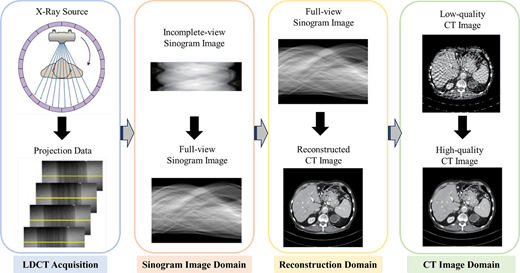
Based on the processing stage, as shown in Figure 2, we introduce a novel taxonomy that categorizes all technologies into two distinct types: image domain processing methods and reconstruction domain processing methods. Based on the shared characteristic of operating directly on image data, the methodologies applied in both the sinogram image domain and the CT image domain can be consolidated under the unified category of image-domain processing techniques. These approaches (Isola et al., 2017; Zhang et al., 2023; Pan et al., 2022) primarily use image processing algorithms to enhance data quality, either by improving the raw sinogram data before reconstruction or by refining the CT images afterward. In contrast, the reconstruction-domain processing technique (Arjovsky et al., 2017; Xia et al., 2022; Lin et al., 2023; Cai et al., 2024; Zha et al., 2024) refer specifically to the core algorithmic process responsible for quantitatively recovering the object’s linear attenuation coefficients (CT values) from the sinogram data, ultimately generating the CT volume (the axial slices of which constitute the final CT images). Section 2 introduces the fundamental principles of CT reconstruction. Subsequently, Sections 3 and 4 present the image-domain processing methods and reconstruction-domain processing methods, respectively. Section 5 introduces the data sets related to CT reconstruction, as well as the evaluation metrics and performance comparisons of different methods. Furthermore, in Section 6, we compare the strengths and limitations of different methods and discuss promising directions for future research.
2. The principle of CT reconstruction
2.1 The process and principle of CT imaging
The typical CT imaging process (Kak and Slaney, 2001; Koetzier et al., 2023; Goldman, 2007) comprises three fundamental stages: projection data acquisition, CT image reconstruction and CT image visualization. These stages, respectively, correspond to:
scanning the subject with a controlled X-ray source and detector array;
computationally solving for the linear attenuation coefficients of tissue voxels from the acquired projection data; and
generating and displaying tomographic images for clinical diagnosis.
2.1.1 Projection data acquisition.
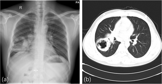
X-rays enable noninvasive visualization of internal structures by penetrating the human body, during which different tissues attenuate the radiation to varying degrees (Brenner and Hall, 2007; Goldman, 2007). In CT imaging, the value associated with each pixel in the resulting image represents the X-ray attenuation coefficient of a volumetric unit (voxel) within a defined tissue slice. Unlike conventional X-ray radiography, which produces a superimposed projection of all tissues along the X-ray path, CT generates true cross-sectional images (Katsuragawa and Doi, 2007; Koetzier et al., 2023). As illustrated in Figure 3, a standard X-ray film displays a composite shadow image of all structures in the beam direction. In contrast, CT provides a series of axial tomographic images, allowing clear visualization of anatomical details layer by layer.
The study (Kak and Slaney, 2001) shows that the intensity of X-rays decays exponentially when passing through a homogeneous substance. represents the original intensity of the X-rays, where the absorption coefficient of each voxel is denoted as and d is the thickness of the voxels. After passing through these voxels, the intensity of the X-ray can be obtained:
through variation,
a continuous representation of this process is:
where represents the absorption coefficient or intensity of the three-dimensional (3D) point.
During a CT scan, the machine rotates around the body to acquire X-ray measurements from multiple angles (Kak and Slaney, 2001; Goldman, 2007). The detector captures the X-rays that have passed through the tissues and converts them into digital signals. The complete set of these angular projections is referred to as the raw data, which is conventionally organized and displayed in the form of sinogram images or projections (Koetzier et al., 2023).
2.1.2 CT image reconstruction.
CT reconstruction refers to the computational process of calculating the linear attenuation coefficient for each voxel of human tissue from the acquired projection data (Kak and Slaney, 2001; Koetzier et al., 2023; Goldman, 2007). This technique essentially solves an inverse problem: based on a set of X-ray projections measured at different angles, it recovers the spatial distribution of attenuation coefficients to produce a final cross-sectional image. The choice of reconstruction algorithm is a primary factor determining the quality of the resulting CT image (Goldman, 2007).
2.1.3 CT image display.
Currently, although certain software tools have been developed to assist in the diagnostic process, the clinical application of CT images still predominantly relies on interpretation by professional physicians to assess human tissue conditions and identify lesions (Kak and Slaney, 2001). Therefore, after CT images are reconstructed, they must be converted into grayscale images that are suitable for visual interpretation. Both electronic CT images for soft-copy reading and films for hard-copy documentation remain in clinical use, each serving specific diagnostic needs (Koetzier et al., 2023; Goldman, 2007).
2.2 Projection and back-projection process
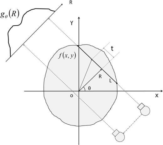
In CT imaging, the scanner rotates around the human body to acquire multi-angle projection data (Kak and Slaney, 2001; Goldman, 2007). It is therefore natural to describe the X-ray projection and back-projection processes in polar coordinates . The projection process is mathematically formulated by the Radon transform (Radon, 2005), while the inverse Radon transform is used in back-projection to recover the distribution of the X-ray attenuation function within human tissues.
As illustrated in Figure 4, the Radon transform (Radon, 2005) converts the Cartesian coordinates (x,y) into polar coordinates , where denotes the attenuation function in Cartesian space. The path L represents the trajectory of an X-ray beam. Then:
the points on the line L are converted to polar representations:
For a parallel X-ray beam at a given angle , the projection function is defined as (Radon, 2005):
3. Image domain processing methods
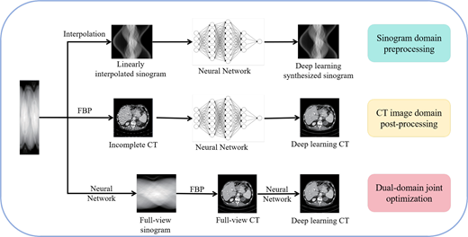
Image domain processing methods work directly on the sparsely sampled sinogram image or the CT image with serious noises and streak artifacts. As shown in Figure 5, these approaches can be classified into three main categories: sinogram domain preprocessing, CT image domain postprocessing and dual-domain joint optimization. Sinogram domain preprocessing methods (Isola et al., 2017; Lee et al., 2017; Fu et al., 2019; Anirudh et al., 2018) aim to convert a incomplete-scan sinogram image into a full-view sinogram image. CT image domain post-processing methods (Zhang et al., 2023; Buades et al., 2005a; 2005b; Geraldo et al., 2016; Liu et al., 2023), on the other hand, reconstruct a CT image from incomplete-scan data and subsequently enhance its quality through operations such as denoising and artifact removal. Dual-domain joint optimization methods (Pan et al., 2022; Zheng et al., 2020; Zhang et al., 2022a; 2022b; Xie et al., 2022) simultaneously refine both the projection data and the reconstructed CT image to ultimately produce a high-quality output.
3.1 Sinogram image domain preprocessing methods
The most immediate problem faced by incomplete-scan CT reconstruction is the lack of part of data in the sinogram image (Lee et al., 2017). One of the most straightforward ways to deal with this is to predict the missing uncollected data according to the incomplete scan sinogram images (Lee et al., 2018; Dong et al., 2019a; 2019b). However, since reconstruction is sensitive to the intrinsic consistency of the sinogram image (Isola et al., 2017; Fu et al., 2019; Bai et al., 2018), care must be taken to avoid artificially introduced artifacts or misinformation when manipulating the sinogram image directly.
The traditional sinogram domain processing method uses an artificially designed interpolation method to estimate the missing neighborhood information from the sparse view of the sine graph to complete the sine map. Simple sinogram diagram interpolation methods include the linear interpolation method (Bertram et al., 2009), the partial differential equation-based method (Kostler et al., 2006) and the frequency domain method (Pohlmann et al., 2014). Improved methods include directional interpolation (Zhang and Sonke, 2013), sine diagram normalized interpolation (Kim et al., 2018) and sine diagram interpolation based on displacement function (Zeng, 2019). These methods can estimate the missing projection data to a certain extent, but as the number of scan angles decreases, the error of the interpolated data gradually increases, thus introducing erroneous data.
With the development of deep learning technology, deep neural networks have shown their powerful capabilities in data completion. With the help of the powerful feature extraction ability of convolutional neural network (CNN) or the generation ability of generative adversarial network (GAN), the sinogram domain interpolation method based on deep learning can recover the complete sinogram graph end-to-end. After obtaining a complete sinogram image, high-quality CT images can be obtained by traditional reconstruction methods. According to the type of neural network, there are two types of interpolation methods, one is CNN-based interpolation method and the other is GAN-based interpolation method.
3.1.1 Sinogram diagram interpolation method based on CNN.
Lee et al. (2017) proposed a CNN-based CT sinogram interpolation model to complete the missing data. The model uses the powerful feature extraction ability of CNN to learn the mapping relationship between sparse view sine diagram and full sine diagram. The neural network is trained by using the interpolated sine diagram and the real complete sine diagram pair, and the model achieves better reconstruction quality than the previous manual interpolation methods. On this basis, Lee et al. (2018) used the residual U-Net to interpolate the sinogram graph, which improved the ability to extract features and obtained a better complete sinogram graph. Similar to (Lee et al., 2017), Dong et al. (2019a; 2019b) combine traditional linear interpolation and residual U-net to complete the sinogram images. Unlike previous works, they experimented on sparse sine patterns that were acquired in real terms instead of synthesizing the data, and reconstructed them with FBP to achieve better CT images.
Then, Dong et al. (2019a; 2019b) coupled the sinogram domain interpolation method based on U-Net and the FBP algorithm, and proposed a new deep learning architecture for reconstructing sparse view sampled CT images. Specifically, the framework first obtains the initial CT image from the sparse view sine diagram by FBP, and then the complete sine image is obtained by projection. The original sine diagram is then input into U-Net for mapping learning, so as to obtain a complete sine diagram. The biggest difference from the previous U-net sinogram diagram interpolation method is that the proposed model uses FBP to initialize the sparse view sine diagram instead of simple linear interpolation initialization. On this basis, Fu et al. (2019) introduced the same deep learning framework to differential phase-contrast computed tomography (DPC-CT), which is a powerful analysis tool for soft tissue and low atomic number samples. They further improved the structure of U-net to extract more features to generate a complete sine map, successfully verifying the excellent performance of this deep learning framework on DPC-CT. In addition, inspired by the super-resolution method of natural images (Tong et al., 2017), Guo et al. (2023) proposed a simplified CNN model for the interpolation of sparse view sinogram images. The model consists of only four convolutional layers, the first three of which are used for feature extraction, and the last layer is a sub-view regression layer. The model can realize the interpolation of the sparse view sine diagram in a short time, and the high-quality CT images can be obtained after FBP reconstruction.
3.1.2 Sinogram domain interpolation method based on GAN.
With the development of deep learning, GANs (Goodfellow et al., 2020) perform well in image inpainting tasks, where effective image information can be added to recover missing parts of an image. The completion problem of a sinogram image is similar to image inpainting, so a imcomplete-scan sinogram image can be done with GAN (Isola et al., 2017; Anirudh et al., 2018; Li et al., 2019a; 2019b). The GAN consists of two parts: a generator and a discriminator, which is used to learn the probability distribution of the training sample and make the recovered image conform to the learned distribution. Discriminators, on the other hand, are used to distinguish whether an image is generated or real. The two modules (Goodfellow et al., 2020) are pitted against each other, allowing the generator to generate a complete sine diagram that approximates the reality.
Anirudh et al. (2018) proposed a 1D and 2D CNN architecture, CT-Net, to achieve the completion of finite-angle sinogram diagrams. The 1D convolutional network is used for the initial interpolation of the sinogram diagram, while the 2D convolutional network repairs the error of the previous step. By generating a near-true complete sine diagram, GAN recovers most of the structural information, allowing for high-quality CT reconstruction. Bai et al. (2018) proposed a generative adversarial network with a joint loss function. In the generator, they trained an autoencoder-like CNN to estimate the missing sinogram domain data, using a discriminator to distinguish between generated and real data. In addition, they used a joint loss function to constrain the generation of the sinogram domain projection map and the quality of the CT image domain. Eventually, they used FBP to complete the CT reconstruction.
Inspired by conditional GAN (cGAN), Li et al. (2019a; 2019b) proposed a sinogram diagram repair model, SI-GAN. The model consists of two parts, an improved U-net generator and a patch design discriminator. After training with sparse sine and full sine pairs, the model can complete the sparse view sine map for CT image reconstruction. However, due to the instability of the GAN, unacceptably false details can sometimes be generated. Therefore, they added an image domain constraint to limit the generation of error information, which is manifested as a weighted image domain loss function. Liu and Li (2020) complete the sine diagram based on a cGAN called Pix2Pix (Isola et al., 2017). Firstly, the sparse view sine graph is initialized by linear interpolation and used as the input of the model. After the complete sine diagram is obtained by the trained Pix2Pix model, the reconstructed CT can be obtained by the FBP algorithm.
3.2 CT image domain post-processing methods
Another strategy of image processing method is to perform post-processing in the CT image domain to enhance reconstructed CT images after sparse angle sampling. Traditional reconstruction methods, such as FBP or SART, were used to reconstruct the sinusoidal pattern sampled from a sparse perspective to obtain the initial CT. Raw CT has artifacts and noise that can affect the diagnosis, so image domain post-processing methods aim to eliminate these artifacts and noise. The idea was natural, and researchers have applied general image processing methods to the enhancement of sparse view CT images. Traditional CT image processing methods use artificially designed fixed convolutional kernels to filter the sparse angle of view to reconstruct CT images, so as to remove artifacts and noise in the images. Classical filtering methods include nonlocal mean filtering (Buades et al., 2005a; 2005b), bilateral filtering (Paris et al., 2009) and statistical algorithm filtering based on maximum posterior estimation (Geraldo et al., 2016). However, due to the nonuniformity of the noise and artifact distribution in sparse view CT images, these artificially designed traditional convolutional filtering methods are difficult to effectively remove image artifacts and noise, and may even cause the image to become more blurry.
With the development of deep learning, a variety of neural network architectures have been proposed to remove artifacts and noise from sparse view CT images. These methods are collectively referred to as sparse view CT image processing methods based on deep learning. After the traditional CT reconstruction method was used to reconstruct the sparse sampling data, the CNN was used to remove artifacts and noise from the images, and finally the high-quality CT images were obtained.
A key paradigm that distinguishes modern deep learning-based reconstruction from pure image-to-image post-processing is the tight integration of the physical imaging model. These physics-aware methods ensure that the final output is consistent with the actual acquired measurements, leading to more reliable and accurate results. Pure post-processing methods operate solely in the image domain. They lack an explicit connection to the raw measurement data. Consequently, they may inadvertently remove or alter anatomically accurate structures while removing artifacts.
Jin et al. (2017) combined residual learning and U-Net to propose a sparse view CT image processing method combining FBP and CNN. After reconstructing the sparse view CT images using FBP, they used the learned residual U-Net to remove artifacts and noise from the CT images, achieving convincing results on both synthetic and real data sets. However, ordinary U-Net is better at processing low-frequency information, resulting in the loss of high-frequency details. Han and Ye (2018) proposed a dual-frame U-Net based on wavelet transform, which adds an additional high-frequency path to the existing U-Net structure, which can better process information in different frequencies. In addition, since the increase in the number of convolutional layers and the increase in the number of channels can easily improve the feature extraction ability of neural networks, larger and deeper U-Nets are also proposed. Mustafa et al. (2020) proposed a multi-channel input/output U-Net CNN model to reconstruct sparse view CT. By using weight-sharing convolution, the model can process a large number of channel data at the same time, only increasing the computational burden of the first and last layers, so as to quickly reconstruct multi-channel high-quality CT images.
By leveraging an adversarial loss, GAN surpasses U-Net in recovering fine image details, thereby effectively mitigating structural blur. Specifically, U-Net only uses a single mean square error to evaluate fidelity, while GAN increases the adversarial training loss on this basis, which can learn the distribution characteristics of the complete CT data, reduce the ambiguity caused by the mean square error, and thus bring the generated reconstruction results. Liao et al. (2018) combined U-Net with GAN to propose a new adversarial learning sparse view CT image post-processing framework. The model uses U-Net as the generator, and designs a new discriminator based on the feature pyramid and differential modulation sword diagram, which greatly eliminates the bar artifacts at various scales through adversarial training. Similarly, Hegazy et al. (2021) used a variant of GAN, U-WGAN, to remove artifacts from CT images. They used a five-stage U-Net in generator and a Wasserstein loss function in the discriminator, which effectively solved the problems of vanishing gradients and unstable training process. Since the artifacts caused by sparse view CT reconstruction are 3D rather than 2D, 3D neural networks have great advantages in the field of CT image post-processing. Xie et al. (2019) proposed a deep encoder-decoder adversarial reconstruction of the 3D network DEAR-3D. By introducing the dense connection module into the generator of GAN, the model can fuse the structural feature information of receptive fields at different levels, and enhance the ability to eliminate artifacts.
More recently, the diffusion model, as a new unsupervised generative model, can produce high-quality images. Liu et al. (2023) introduced it into the post-processing of finite-angle CT reconstruction, combined with the denoising diffusion probabilistic model (Ho et al., 2020), and proposed the first ensemble conditional training diffusion model and explicit physical measurement model DOLCE. The model consists of two parts: a fixed forward process and a learning-based reverse process. By inputting a finite-angle CT image during the reverse diffusion process, DOLCE can stably generate a CT image with artifact removed. Notably, DOLCE operates in an unpaired training manner and leverages a conditioning mechanism to guide the reverse diffusion process. Despite a higher run-time cost compared to deterministic methods, this model provides substantial post-processing benefits by generating high-quality, artifact-free images.
In addition, Transformer can use its multi-head self-attention mechanism to extract global information, which also plays an important role in sparse view CT image processing. Zhang et al. (2023) combined CNN and Transformer to propose a new encoder model, TIME-Net, for CT image processing with limited viewing angles. The model consists of three encoders, two CNN encoders are used to extract high-level features and capture structural information, and the Transformer encoder is used to capture global information. The features extracted by the three encoders are fused and compressed, and high-quality CT images can be generated by CNN decoders. However, it is worth noting that the addition of attention mechanisms makes the processing process more computationally expensive and time-consuming.
3.3 Dual-domain joint optimization methods
The previous two kinds of methods are to interpolate or filter on a single domain to achieve the optimization of a single domain, which often cannot take into account the processing effect on the other domain. To further reduce the error caused by sparse sinogram image interpolation and the artifact noise of CT images, various dual-domain joint optimizations are proposed. The complete sinogram image is obtained by adaptive learning on the incomplete sinogram image, and then the CT image is obtained by traditional reconstruction methods such as FBP, and the image post-processing is carried out in the CT image domain. dual-domain joint processing methods allow the model to constrain each other between the two domains, so that high-quality CT images can be generated end-to-end.
Zheng et al. (2020) constructed a dual-domain deep learning framework with three modules for the sparse view CT reconstruction of spiral CT. Firstly, the projection domain CNN is used to estimate the missing projection data, then it is converted into 2D sector beam projection data, and the projection data is reconstructed into CT images by FBP algorithm, and finally, the CT image domain CNN is used to optimize the reconstructed images. The framework realizes the end-to-end sparse view spiral CT reconstruction, and verifies its effectiveness on the real data sets. Hu et al. (2020) proposed a mixed domain neural network, HDNet, using two 3D U-Nets combined with the FDK algorithm. The model divides the process of sparse view CT reconstruction into two stages, namely, sine domain data completion and post-processing of CT image domain. The neural network of the two stages is connected by FDK’s reconstruction of the sinogram image to the CT image. The phased learning method greatly reduces the complexity of reconstruction, and the parallel processing of mixed domains also greatly improves the detail preservation and overall quality of the image. Zhou et al. (2022) proposed a recursive cyclic dual-domain CT reconstruction framework, DuDoDR-Net, to reconstruct artifact-free CT images through the restoration of cyclic image domain and sinogram domain. In addition, they have developed an image Data Consistency Layer (iDCL) and a sinogram graph data consistency layer (sDCL) in a recursive framework to ensure the quality of the reconstruction. Zhang et al. (2022a; 2022b) extended the state-of-the-art LEARN model to a dual-domain optimized version and proposed the LEARN++ model to reconstruct sparse view CT. The model is composed of two parallel and interactive CNN sub-networks, which realize the completion of sinogram graphs and the post-processing of CT images, respectively. The model is jointly optimized in the sinogram domain and CT image domain, and makes use of the latent relationship between the dual-domain data, and achieves more advanced results in both artifact removal and detail information preservation.
Like the previous single-domain processing methods, the GAN model is also introduced into the dual-domain joint optimization method for sparse view CT reconstruction. Zhao et al. (2018) combined CNN and GAN to propose a novel framework for dual-domain joint optimization to generate high-quality CT images. Specifically, they trained a CNN with an MSE loss function to complete the sinogram image, reconstructed the except CT images using FBP or WHLS methods, and finally trained a GAN with a perceived loss function to process the reconstructed CT images to obtain a high-quality final output. The joint optimization method changed the shortcomings of the single-domain processing method resulting in smooth and blurry images, and obtained CT images with more detailed information. Wu et al. (2021) proposed an optimization network based on dual-domain residuals, DRONE. The model is composed of three modules with different functions, namely embedding, optimization, and perception modules, which realize the initialization, refinement, and structural perception of CT image reconstruction on dual domains. Similarly, Ketola et al. (2021) proposed a new dual-domain joint optimization model, DGAN, using two GANs for sparse view CT reconstruction. The generators and discriminators of the two GAN models are U-Net and PatchGAN, respectively. The two GAN models, one is used to complete incomplete sinogram images, and the other is used to process the CT images reconstructed by FBP. At the same time, they added real sine images and CT images to the training process to suppress false information generation and improve the detailed structure of the images. On this basis, Xie et al. (2022) used an unsupervised GAN in the CT image post-processing domain to reduce the dependence on label data, and used another GAN to provide a complete sinogram map for the FBP algorithm.
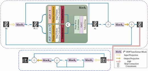
Due to the learning ability of the Transformer model for long-distance dependence, it is also used to construct a dual-domain joint optimization model in combination with FBP. To solve the problem of sparse view CT, Shi et al. (2022) proposed a Transformer-based dual-domain CT reconstruction network model. The model treats the sparse sinogram diagram as a long sentence in NLP to assist the Transformer encoder in generating high-quality CT images. As shown in Figure 6, Li et al. (2022) proposed a parallel Transformer dual-domain CT reconstruction network, DDPTransformer, to reconstruct sparse view CT. The model consists of four stages, namely, sinogram image interpolation, sinogram domain processing, FBP and CT image domain processing. At the same time, considering that the processed image is 2D, they replace the multilayer perceptron (MLP) in the Transformer with layer-convolution-layer to enhance the feature extraction ability. Pan et al. (2022) proposed a Multi-domain Integrated Swin Transformer network to improve the quality of CT images. The model consists of three stages, the first stage was to extract image features in the projection domain and the CT image domain, the second was to detect and protect the edge of the image, and the last stage is to enhance the processing module of the CT image. The difference is that the first two stages are composed of CNNs, and the last module is composed of Transformer to extract global features. This design greatly reduces the computational overhead and time complexity caused by dual-domain joint optimization.
4. Reconstruction domain processing methods
Reconstruction-domain methods refer to techniques that generate CT images directly from incomplete-scan projection data. Traditional algorithms in this domain, such as filtered back-projection (Shepp and Logan, 1974) and IR (Andersen and Kak, 1984), often produce severe streaking artifacts and noise when applied to incomplete-scan data. The advent of deep learning has progressively overcome this limitation (Arjovsky et al., 2017; Xia et al., 2022; Lin et al., 2023; Cai et al., 2024; Zha et al., 2024; Zhu et al., 2018). Consequently, reconstruction domain approaches have evolved from deep learning-enhanced analytical (Wurfl et al., 2018; He et al., 2020) or iterative methods (Sauer and Bouman, 1993; Buades et al., 2005a; 2005b) to fully end-to-end deep learning mapping techniques (Lin et al., 2023; Zang et al., 2021; Li et al., 2023).
4.1 Combination of traditional CT reconstruction algorithms and deep learning
The traditional CT reconstruction algorithms mainly include analysis methods (Shepp and Logan, 1974) and iterative methods (Andersen and Kak, 1984). The analysis methods mainly include filtered back projection algorithms (Shepp and Logan, 1974) and back projection analysis methods based on deep learning (He et al., 2020; Wurfl et al., 2018; Xie et al., 2020). The main development of iterative methods consists of three stages: the traditional IR algorithms (e.g. SART (Andersen and Kak, 1984)), the second stage is the IR algorithm based on model optimization (Chen et al., 2015; Huang et al., 2018; Xu et al., 2020), and the third stage is the IR algorithm based on deep learning. Deep learning technology (LeCun et al., 2015) can make up for the shortcoming of previous methods. when combined with it, the quality of reconstruction can be greatly improved (Zha et al., 2022; Shen et al., 2022).
4.1.1 Analytical methods.
The analytical method estimates the absorption coefficient by solving the Radon transform and its inverse equation. The common methods are to filter back projection (FBP) (Shepp and Logan, 1974) and its 3D conical beam variant, FDK (Feldkamp et al., 1984). These methods provide analytical reconstruction based on the Radon transform and its inverse transformation. The success of these transformation-based methods depends on their fast reconstruction speed, as well as providing reasonable quality results when a sufficient number of projections are available. With the advantages of high spatial resolution, these methods were widely used in early CT instruments. However, in the case of sparse angle sampling, the CT images obtained by such a method have a low signal-to-noise ratio and have severe streak artifacts.
The deep learning-based back-projection algorithm (Cai et al., 2020; Zha et al., 2024; Goldman, 2007; Radon, 2005; Lee et al., 2017; Fu et al., 2019) combine the advantages of deep learning and back-projection algorithms. Using a data-driven approach and the physically guided process of the back-projection method, these methods not only achieve higher reconstruction quality, but also reconstruct faster than general neural network methods.
Wurfl et al. (2018) mapped the back-projection algorithm to the neural network, reducing artifacts caused by the absence of projection data. They designed a unique back-projection layer in place of the fully connected layer (FCL), which reduced memory overhead. At the same time, the neural network model can automatically learn the weight value of the projection domain in the back-projection algorithm, and the back-projection layer allows the joint optimization of the projection domain and the CT image domain. This provides a good balance between reconstruction performance and speed. However, the algorithm only demonstrates the advantages of learning projection domain weights. Li et al. (2019a; 2019b) also designed a neural network architecture iCT-Net based on the principle of FBP, which reconstructs CT images from sparse view sinogram images. This neural network first denoises the sinogram domain data, and then designs a back-projection layer with learnable weights through the convolutional layer and the FCL to achieve CT reconstruction. He et al. (2020) proposed a deep learning-based reconstruction architecture of Jordan inverse, iRadonMAP. This neural network contains three specialized components. The first component is a fully connected filter layer in the sinogram domain along the direction of the rotation angle, the second component is the sinogram domain back-projection layer, which backprojects the sinogram domain data filtered by the first layer into the spatial domain, and the third is a general-purpose CNN to improve the quality of the reconstruction and the overall performance. iRadonMAP achieved better reconstruction quality with a learnable FBP algorithm.
On this basis, He et al. (2021) proposed a lightweight model DSigNet, which uses complete prior knowledge to convert the real projection-CT mapping relationship into a virtual relationship in a data-driven way. This virtual imaging system uses a lightweight back-projection module and a downsampled geometry modeling module for faster reconstruction. Xie et al. (2020) also proposed a neural network architecture, DEER, based on the principle of FBP to realize CT reconstruction with sparse view. DEER consists of two parts: a generator network and a discriminator network. The generative network consists of two modules: backprojection and optimization. Since each point in the sine diagram is only related to the pixel value on the relevant X-ray path through the CT image, the other pixels do not contribute much to this. DEER uses the FCL to learn the global structure point by point, so as to learn the parameters of the reverse projection process.
4.1.2 Iterative reconstruction methods.
The IR algorithm refers to the method of iteratively reconstructing CT images through the process of repeated projection and back-projection many times. The classic IR algorithms include ART (Gordon et al., 1970) and SART (Andersen and Kak, 1984). Sauer and Bouman (1993) proposed a Bayesian iterative method that updates individual pixels rather than the entire image during each iteration, with better convergence in a high spatial frequency mode. Manglos et al. (1995) proposed a new CBCT IR method based on Ordered Subset Expectation Maximization, and obtained high-quality IR images. Pan et al. (2006) improved the ART and SART algorithms, and proposed two variable weight IR algorithms (VW-ART and VW-OS-SART). In both algorithms, the weights vary with the geometric direction of the rays, and a better reconstruction quality is successfully achieved. Compared with the analysis method, the IR algorithm can achieve better quality in the sparse view CT reconstruction task, but there are still structural blurs and artifacts.
To overcome the above problems, researchers have introduced prior information into IR algorithms (Buades et al., 2005a; 2005b; Chen et al., 2015; Huang et al., 2018; Xu et al., 2020; Sidky and Pan, 2008; Zang et al., 2018). These methods are called model-based iterative reconstruction (MBIR). Based on the development of compressive sensing theory and sparsely sampled image reconstruction technology (Candes et al., 2006), Sidky and Pan (2008) developed a new iterative algorithm ASD-POCS. The algorithm introduces TV regularization, uses convex set projection (POCS) to enforce constraints, and minimizes TV targets by the steepest descent method with adaptive step size. Chen et al. (2015) introduced the reconstruction method of non-local mean (NLM) (Buades et al., 2005a; 2005b), and proposed a model for sparse projection image reconstruction, ART-ANLM, which balances the relationship between structural information retention and noise suppression. To solve the problem of multi-scale artifacts, Huang et al. (2018) proposed two different scale spatial anisotropy TV (ssaTV) algorithms, which can reduce the existence of artifacts more efficiently. Zang et al. (2018) designed a new 3D structural tensor prior and incorporated it as a regularizer in the iterative optimization method for CT reconstruction. Xu et al. (2020) combined dictionary learning (DL) and image gradient L0-norm into an image reconstruction model. Dictionary learning can improve the preservation of image detail information, while image gradient L0-norm can reduce artifacts and further improve the image quality of sparse view CT reconstruction.
MBIR has successfully improved the reconstruction quality of CT images by introducing various regular terms, but there are two significant problems:
The addition of regular terms makes the computational complexity very large, and the reconstruction process requires a large amount of memory and a longer time.
These regular terms and hyperparameters usually require manual selection to deal with different systems and reconstruction scenarios, which requires the participation of specialized human knowledge, and also reduces the generalization ability of different fields.
However, the IR method based on deep learning (DLIR) can overcome these difficulties to a certain extent. By training a neural network to replace the manual selection of regular terms and hyperparameters, the efficiency and generalization ability of the iterative model can be greatly improved.
To solve the above two problems of the iterative model, Chen et al. (2018) constructed a neural network model based on the evaluation of learning experts (LEARN). The model uses a fixed number of iterations, and the process is expanded into a neural network, which allows the parameters and regularization terms to be trained using external data sets, thus enabling adaptive learning of parameters and regularization terms. Hammernik et al. (2017) used CNN instead of the convolutional operator regular term to iteratively update the parameters, and at the same time, the equilibrium parameters were adaptively learned through backpropagation, which reduced the computational complexity of the iterative model. Similarly, to avoid the choice of regularization terms and regularization parameters, Wang et al. (2019) proposed a deep reconstruction (ADMMBDR) algorithm based on the alternating direction multiplier method (ADMM). They used the ADMM algorithm to decompose a regularization reconstruction model, and then replaced a portion of the ADMM algorithm with a deep CNN to reduce artifacts and adaptively learn regex and balance parameters.
Bubba et al. (2019) developed a hybrid reconstruction framework based on deep learning that combines model-based sparse regularization and data-driven. The model resolves the wavefront set in phase space through the shear wave transform, learning only the parts that the model-based approach cannot handle, and the neural network can infer unknown shear wave coefficients. Compared with model-based or data-based reconstruction methods alone, the hybrid model achieves better reconstruction results. Iterative neural network (INN) combines regression neural network and iterative MBIR algorithm, which can bring good generalization ability and better reconstruction quality, and is a better iterative reconstruction method (DLIR) for deep learning. Chun et al. (2020) proposed the first fast-converging INN model, Momentum-Net. The model consists of three core modules: image refining, extrapolation and MBIR. By using momentum updates to speed up, Momentum-Net has faster convergence and higher reconstruction quality. Xiang et al. (2021) combined the advantages of a model-based rapid iterative shrinkage/threshold algorithm (FISTA) and a data-driven neural network to propose FISTA-Net. The model expands the iterative steps into cascading blocks, and develops a near-end operator network for nonlinear thresholding. Key parameters can be learned from the training data rather than manually selected, and FISTA-Net shows good generalization ability.
Xia et al. (2022) combined Transformer and CNN into the CT IR algorithm, and proposed a learning-based local-non-local regularization model, RegFormer, to reconstruct sparse view CT images. The model unfolds the iterative scheme into a neural network and adaptively learns regularized terms. RegFormer uses CNN to learn local information of images, and uses the self-attention encoder structure in Transformer to learn nonlocal prior information, which improves the ability to extract deep features in the iterative process. The model achieves competitive performance in both artifact removal and detail preservation.
4.2 End-to-end incomplete-scan CT reconstruction algorithms based on deep learning
CT image reconstruction is often formulated as an inverse problem, which can be mathematically expressed as the inverse of the projection process (Chung et al., 2023):
In the formula, y denotes the sinogram field projection data; x is the CT image vector; A is the projection matrix, which corresponds to the projection process; e indicates noise or other uncontrollable influencing factors during CT projection. The process of CT reconstruction is the process of finding x from y in reverse. Under the condition of sparse projection perspective, deep learning-based technology has made good progress in solving such inverse problems. In particular, the deep learning method of end-to-end mapping does not require basic knowledge of medical imaging to directly learn the mapping relationship between projection map and CT image. According to different data-driven approaches, this review divides end-to-end CT reconstruction into two categories: sparse view CT reconstruction based on supervised learning and sparse view CT reconstruction based on self-supervised learning.
4.2.1 Incomplete-scan CT reconstruction algorithms based on supervised learning.
The supervised learning-based approach uses a data-driven model to learn the mapping relationship between projected and CT images. The training process often requires a large number of projection-CT data pairs, and after the training is completed, the trained model can realize the end-to-end mapping reconstruction of the projected image to the CT image.
Zhu et al. (2018) first proposed a unified multi-domain image reconstruction framework, AUTOMAP. It replaces the previously hand-crafted signal processing sequence module and allows you to learn the mapping between sensors and image domains without specialized knowledge. Specifically, AUTOMAP uses CNNs to construct a deep neural network, using three FCLs followed by two convolutional layers to build the framework. This technique uses data-driven supervised learning to learn the mapping between the sensor domain and the image domain. Compared with traditional manual reconstruction methods, the noise resistance is stronger and there are fewer reconstruction artifacts. On this basis, Zhang and Liang (2020) studied the role of FCLs in the sinogram domain to image domain method. Through the visual analysis of the weights of the FCL, they found that the main function of the FCL is to realize the back-projection function in the process of CT reconstruction. This finding explains the role of various parts of the end-to-end learning framework and plays an important role in the development of deep learning-based CT reconstruction technology. Yim et al. (2021) reduced the dimension of the FCL to equal to the dimension of the image expansion, and increased the number of convolutional layers. Compared with traditional reconstruction methods in various indicators, this model can provide high-quality CT images.
To optimize the time and space overhead of the FCL, Ma et al. (2020) used a series of smaller FCL to replace the larger FCLs based on the sparse non-negative matrix factorization theory on the AUTOMAP framework. At the mathematical level, the projection matrix is approximately decomposed into a series of low-dimensional matrices by using NMF by using the non-negative properties of the projection matrix values. At the same time, a multiscale feature fusion U-net is added behind the FCL to further improve the quality of the reconstructed image. The decomposition of the inverse projection matrix has been verified in both simulation and practical experiments, and its effectiveness has been proved. The method of using a fully convolutional network instead of a FCL for reconstruction can greatly reduce the time of CT reconstruction without sacrificing the quality of CT reconstruction. Kalare and Bajpai (2020) proposed a deep reconstruction network based on cascaded U-net RecDNN, which uses a fully convolutional network to perform CT reconstruction on projected images. Mizusawa et al. (2021) proposed an end-to-end reconstruction network based on stacked U-net. Due to patient privacy concerns, there are few training images for medical images, so it is not easy to create highly accurate models. As a result, they also utilized a variety of images from ImageNet, a visual database.
Although researchers have been working to reduce the number of CT scan angles, ultra-sparsely sampled tomography is difficult to achieve. On the other hand, Shen et al. (2019) and Ying et al. (2019) use a single projection image or two X-rays to reconstruct the CT volume. Both are designed with a converter to convert 2D features into 3D features for CT reconstruction. Shen et al. (2019) designed a hierarchical ultra-sparsely sampled CT reconstruction network. The first-layer network extracts the 2D features of the X-ray film, the second-layer network realizes the conversion of 2D features to 3D features, and the third-layer network realizes the generation of CT volumetric images. The network is trained on sampled CT images from different perspectives of specific patients, and a single X-projection image can be used to generate CT volume images after training. Based on deep learning, Ying et al. (2019) proposed the first model X2CT-GAN for CT reconstruction from biplanar X-ray images. The model proposes a new jump connection module to bridge 2D to 3D features, which has not been covered by previous GANs, and achieves good results. It is worth noting that although this model belongs to the supervised learning CT reconstruction method, unlike the previous models, X2CT-GAN uses two orthogonal X-ray light sheets instead of a projection map in the sinogram domain. Although it cannot be compared with the effect of CT instruments, it greatly broadens the idea of sparse view CT reconstruction.
The above methods can only estimate CT volume and require another challenging segmentation step to extract a 3D reconstruction of the dissected object. Kasten et al. (2020) proposed a novel end-to-end deep learning method for reconstructing the knee joint from two biplanar X-ray images. This method adopts a new 3D information representation and retains the two-view collection constraint. This new CNN architecture can generate accurate CT knee 3D structures. Previous sparse view CT reconstruction methods based on supervised learning are all voxel-based techniques, Lin et al. (2023) proposed a point-based model DIF-Net. It should be noted that although the model is measured in points, it is different from the NeRF-based technique in the following self-supervised learning methods. DIF-Net does not use neural rendering, but adopts the methods of extracting features, fusing features and estimating density, and still needs projection-CT image pairs for training. The model designs an encoder to extract the features of the projected image of each view, perform feature fusion in points, estimate the density of the point and compare it with the real density value for optimization. Compared to voxel-based supervised learning methods, DIF-Net requires fewer projections, is faster to reconstruct and is of higher quality.
4.2.2 Incomplete-scan CT reconstruction algorithms based on self-supervised learning.
Most powerful models based on supervised learning methods, such as CNNs (He et al., 2016), learn direct mapping from 2D X-ray projection maps to CT volumes. One of the main drawbacks of these deep learning methods is that network training requires a large number of projection-CT pairs, which are difficult to obtain in practice. Collecting even a small projected CT data set is tedious, and harmful to health. In addition, because the statistic characteristics of CT data are domain-specific, domains vary widely between different CT data sets, making it difficult to transfer network learning from one application area to another. Compared with the algorithm based on paired learning, the self-supervised learning method does not require CT volume for training in tomography reconstruction. Instead, they only need projections of a scene. Unlike the relational learning of projection and CT volume, self-supervised learning learns the contiguous space as a specific property and generates a projection map through the rendering process. By optimizing this attribute via the sparse-view projection map, accurate 3D information can be reconstructed to generate CT volumes. In this review, self-supervised learning methods were divided into two categories, namely, incomplete-scan CT reconstruction based on implicit neural representation and incomplete-scan CT reconstruction based on 3D Gaussian.
4.2.2.1 Incomplete-scan
CT reconstruction algorithms based on implicit neural representation. Mildenhall et al. (2021) proposed Neural Radiance Field (NeRF) based on continuous implicit representation, which successfully produced high-quality results in Novel View Synthesis tasks. They represented the scene with a fully connected network and optimized the Neural Radiance Field to synthesize a new view of the scene with complex geometry and appearance. The advent of NeRF also provides more reasonable solutions for X-ray 3D reconstruction. In this approach, a MLP network is used to represent the density field of the scanned object as a function of the input coordinates. This network is then learned from the captured projections. This representation provides greater flexibility to produce a composite projection of any desired resolution.
However, although RGB-NeRF algorithms have been well developed, applying them directly to X-ray scenes may yield suboptimal results due to the fundamental differences between RGB and X-ray imaging. Standard Structure from Motion (SfM) is unsuitable for CT as X-ray projections lack the features needed for pose estimation. Hence, scanner geometry is typically pre-calibrated.
NeAT (Ruckert et al., 2022) introduces a pioneering deep learning rendering framework tailored for tomography. It uses an adaptive multilevel representation based on an explicit octree structure. This design facilitates efficient empty space skipping and adaptive sampling concentration in regions of high complexity, thereby improving computational efficiency. In addition, the incorporated neural features can serve as an effective regularizer for better reconstruction. Experimental results demonstrate that NeAT achieves superior reconstruction quality compared to conventional CT reconstruction techniques. This is also the first successful application of INR in a tomography reconstruction task. At the same time, the researchers found that the introduction of other factors into the INR resulted in higher reconstruction quality. NeRF successfully reconstructs scenes with high-frequency detail by utilizing positional encoding and retargeting conditions. Tancik et al. (2021, 2020) generalized the concept of positional coding to more general Fourier features. Sitzmann et al. (2020) demonstrated that the periodic activation function helps to represent complex signals and their derivatives of objection in fine detail. Zang et al. (2021) combined the advantages of implicit neural representation and prior information to propose the IntraTomo framework to solve the problem of pathological inversion in sparse view CT reconstruction. The model consists of two core modules: a coordinate-based 3D reconstruction mapping network and a geometric optimization module. In the first module, the unknown density field is represented as a continuously differentiable function, parameterized by a neural network. Specifically, they trained an MLP with the input as encoded spatial coordinates and the output as the density of the location.
At the same time, to overcome this bias, IntraTomo introduced the Fourier feature (Tancik et al., 2020) into the network mapping module because the study (Rahaman et al., 2019) found that MLP could not recover the high-frequency details of the target. This neural network is optimized in a self-supervised learning manner by projection maps sparsely sampled by CT instruments. In the second module, the density field is consistently refined by a local and non-local geometric prior information. Through the optimization of these two modules, IntraTomo has achieved unprecedented success in sparse view CT synthesis tasks, and obtained qualitative and quantitative results on different data sets that are ahead of the previous methods. However, although the Fourier frequency domain coding method has achieved better reconstruction results, the speed of INR-based CT reconstruction is still slow. Zha et al. (2022) proposed a new NeRF-based framework (NAF), which is also a self-supervised solution for sparse view CT reconstruction. They used INR to parameterize the density field and simulated the attenuation process of X-rays with a new-view picture rendering process from a self-supervised network. The difference is that NAF does not use the common Fourier frequency domain coding, but uses hash encoding (Muller et al., 2022), which reduces the number of parameters of MLP. The neural network training uses the sampled sparse view projection map to self-supervised training, and after the training is completed, the density value of the CT volume is obtained by direct summation to generate the corresponding CT image. The NAF model takes advantage of the smoothness and sparsity of human organs to achieve more advanced results and, most importantly, it takes only a fairly short time compared to IntraTomo.
In clinical practice, patients sometimes have multiple CT scans over time, and the use of previous CT images to provide information is an effective way to reduce the dose of CT radiation. Based on this application scenario, Shen et al. (2022) proposed the method of NeRP. Unlike previous deep learning-based image reconstruction methods, NeRP reconstructs CT images using internal information from prior images and sparsely sampled projection features. Specifically, NeRP uses implicit neural representation to realize the mapping relationship learning from 3D coordinates to density values. The difference is that before optimizing the density field with a sparse projection image, NeRP embeds the information of the prior image into the neural network. When optimizing with a sparse view projection image, the neural network only needs to change the area that is minimal in a short period of time. As a result, NeRP can take advantage of a sufficiently small projection map to capture this subtle but important variation. In summary, NeRP first uses longitudinal CT images to embed most of the density field information, and then captures the changing structure through sparsely sampled projection maps, so as to achieve high reconstruction quality and greatly reduce the radiation dose of patients, laying a solid foundation for potential clinical applications.
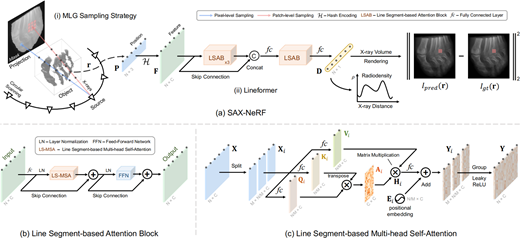
Existing NeRF algorithms treat every spatial point equally, while ignoring the correlation between different points on X-rays, which leads to the limitation of structure capture. Considering this characteristic, Cai et al. (2024) proposed a perception framework for sparse view CT reconstruction SAX-NeRF, which is also an implicit neural representation method for mapping 3D space as a continuous density field. The difference is that the framework is the first exploration to use Transformer for X-ray neural rendering. As shown in Figure 7, SAX-NeRF designed a line-segment-based converter as the core, and accurately captured the internal structure information of the 3D object by modeling the dependencies within each line segment of the X-ray. In addition, the method of INR usually adopts a pixel-level X-ray random sampling strategy. As a result, the contextual structure information in the 2D projection diagram is not well utilized. The random sampling method will also cause the empty area to calculate, which greatly reduces the efficiency of reconstruction. Therefore, SAX-NeRF proposes an MLG ray sampling strategy, which uses masks to segment the foreground of the image, excludes blank areas, and uses pixel-level and block-level sampling methods to help transformer module perceive the local context structure information. Sax-NeRf introduced transformer into the neural representation of X-rays, and achieved more advanced reconstruction results on multiple data sets.
4.2.2.2 Incomplete-scan. CT reconstruction algorithms based on 3D Gaussian
Although the sparse-view CT reconstruction method based on neural radiance field (Mildenhall et al., 2021) has achieved good reconstruction quality, due to the mapping based on 3D points, a large number of point samples are required in the training and rendering process, which is very time-consuming. This is also contrary to the time-sensitive requirements of medical diagnosis, and the results cannot be provided in time, which leads to the difficulty of practical application of the algorithm. The 3D Gaussian splatting model (Kerbl et al., 2023) has recently revolutionized this situation, using highly parallel rasterization for image rendering, which is much faster than the NeRF method.
Just as the way that CT reconstruction is introduced to NeRF (Mildenhall et al., 2021), 3D Gaussian model is first directly applied to the task of composing novel X-ray views (Cai et al., 2025; Nikolakakis et al., 2024), and then to the task of CT reconstruction (Zha et al., 2024). Training a 3D Gaussian-based representation of the scene is much faster, which is critical in medical imaging.
Nikolakakis et al. (2024) used the Gaussian splatting framework to construct the model GaSpCT, and realized the synthesis of a novel perspective based on finite 2D projection images. And it does not require the structure of motion (SfM) method in Schonberger and Frahm (2016) to initialize the Gaussian element position with prior information. In addition, due to the natural smoothness of tissues and organs, the model strengthens the baseline loss function and introduces the total variational loss and the negative log-likelihood loss of the beta distribution, making it more suitable for X-ray imaging use cases, which is also applicable in the task of generating CT images from CT reconstruction. The sparse view sampling feature of the model reduces the total scan time and the radiation dose received by the patient, and greatly improves the speed of reconstruction and rendering. Cai et al. (2025) also built the X-Gaussian model based on the 3D Gaussian framework for the task of generating novel X-ray views. Taking advantage of the anisotropy of X-ray imaging, they designed a novel radiation Gaussian point cloud model and rasterizer, and customized an ACUI initialization strategy to make the model more suitable for CT reconstruction and 2D projection imaging tasks. Significant savings in the speed of reconstruction and rendering, while also achieving more advanced quality. Although the tasks of these two works are different, they have greatly promoted the application of Gaussian Splatting in the task of sparse view CT reconstruction, but unfortunately, they are only one step away. Zha et al. (2024) proposed the first Gaussian splatting based sparse view CT reconstruction framework, , which highlights its important practical value in sparse view CT reconstruction tasks due to its excellent performance and fast training.
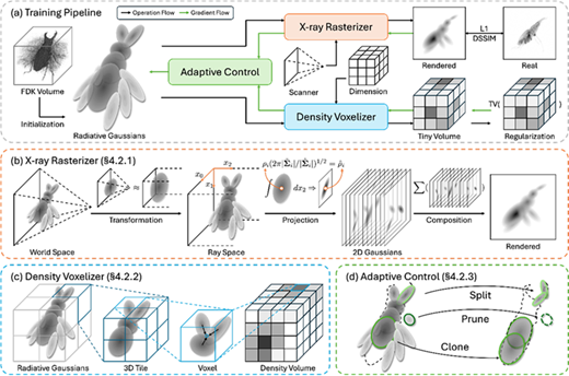
As shown in Figure 8, model first introduces a custom Gaussian kernel. Specifically, the model discards two properties of opacity and spherical harmonic sparsity in the original 3D Gaussian model, and replaces them with the absorption intensity corresponding to the CT value. It is worth noting that the intensity value is directly multiplied with 3D Gaussian as a representation of the Gaussian kernel, and the attribute kernel density of the Gaussian kernel is quickly optimized through the CT projection image of the sparse view. Different from the task of novel perspective synthesis, the ultimate goal of this model is to obtain reconstructed CT images, so they develop a CUDA-based differentiable voxelizer modeled after the rasterizer of the rendering process to obtain CT images. Gradient optimization of Gaussian unwrapping requires the camera pose of the input image in the 3D scene. X-ray images and CT images are both grayscale images with lower contrast compared to RGB images. Also, due to the transmitted imaging nature of X-rays, different layers of the object overlap in the same location of the projection. These two issues reduce the accuracy of feature detection and matching in SfM. Therefore, the SfM method is not suitable for CT reconstruction tasks. Like in Refs (Cai et al., 2025; Nikolakakis et al., 2024), model does not use the SfM method to initialize the point cloud and obtain the camera parameters, but initializes the radiative Gaussian distribution through the preliminary results obtained from the analytical method FDK (Feldkamp et al., 1984). The camera internal and external parameters are generated directly from prior knowledge of the CT scan parameters provided by DICOM metadata. Verified on multiple data sets, model can obtain high-quality results within three minutes, which is more in line with the time requirements of medical application scenarios, which greatly promotes the development and application of sparse view CT reconstruction algorithms.
5. Data sets and metrics
5.1 Data sets for CT reconstruction
For CT reconstruction, AAPM Low Dose CT (McCollough et al., 2017), LIDC-IDRI (Armato et al., 2011), TCIA (Clark et al., 2013), XCAT phantom (Segars et al., 2008) and Shepp-Logan phantom (Shepp and Logan, 1974) data sets are five widely used public data sets.
AAPM Low Dose CT data set (McCollough et al., 2017) primarily consists of abdominal CT scans from 10 patients, providing paired normal-dose CT images and the raw projection data. Quarter-dose and low-dose CT sinograms are simulated by inserting Poisson noise into the original projections to mimic the noise level of a 75% dose reduction, creating a benchmark for developing and evaluating denoising and reconstruction algorithms by offering ideal low-quality input and high-quality reference data pairs. The projection data is stored in an extended vendor-neutral DICOM format (DICOM-CT-PD), which includes the acquisition geometry, facilitating research in both the image and projection domains.
LIDC-IDRI data set (Armato et al., 2011) is a major public reference database for lung nodule detection and classification research, comprising 1,018 low-dose chest CT scans from 1,010 patients. It is distinguished by its detailed annotations of 7,371 lung nodules, which include precise markings of the centroid, boundaries and morphological characteristics.
TCIA (The Cancer Imaging Archive) (Clark et al., 2013) is a large-scale, open-access platform funded by the National Cancer Institute that serves as a public repository for medical images related to cancer, hosting curated collections of de-identified clinical images in DICOM format, such as CT, MRI and PET scans.
XCAT phantom (Segars et al., 2008) is a high-fidelity, whole-body computational model based on real human anatomical data, which utilizes Non-Uniform Rational B-Splines surfaces to define thousands of detailed anatomical structures for both male and female models. This phantom can produce synthetic image data for various modalities like CT, MRI and PET.
Shepp-Logan phantom (Shepp and Logan, 1974) is a standard digital test image data set widely used in the field of medical imaging. It consists of a large ellipse, which contains several smaller ellipses of varying sizes, intensities and orientations that simulate internal features of the brain. This mathematical model is valued for its precisely defined structure, which provides a known ground truth for quantitatively evaluating the performance of image reconstruction algorithms.
5.2 Metrics
The quality of CT images is directly related to the difficulty of clinical diagnosis, so any CT reconstruction method needs to evaluate its image quality. A good CT image must be authentic and reliable. Common objective evaluation indexes of CT images (Sara et al., 2019) include signal-to-noise ratio, mean square error, peak signal-to-noise ratio, structural similarity index method and feature similarity index method.
PSNR (peak signal-to-noise ratio) describes the ratio between the maximum possible power of a signal and the power of the destructive noise that affects the accuracy of the signal’s representation. Because many signals have a very wide dynamic range, PSNR is often expressed in logarithmic decibel (dB) units. The decibel unit is a more intuitive reflection of the relative strength between the signal and the noise. The higher PSNR value means the higher quality of the CT image (Hore and Ziou, 2010). It can be calculated by the following formula (MSE is the mean square error and MAX is the maximum power of the signal):
SSIM (structural similarity index method) is a measure of the similarity between two images. This metric models distortion as a combination of three attributes: brightness, contrast, and structure. Brightness is estimated using the mean, contrast is estimated using the standard deviation, and structural similarity is estimated using covariance. In practice, the value of SSIM is usually between 0 and 1, with closer to 1 indicating higher image quality, and closer to 0 indicating worse image quality (Wang et al., 2004). The formula for calculating SSIM is as follows:
here, x and y are the two images, and are their mean, and are their standard deviation and means their covariance. and are constants set to avoid a zero denominator, and are usually taken as , . L is the dynamic range of pixel values (e.g. for an 8-bit image, L = 255), and are small constants, usually taken as = 0.01 and = 0.03.
FSIM (feature similarity index measure) measures the similarity or quality difference between two images. Based on the feature information of the image, the image quality (Zhang et al., 2011) is evaluated by calculating the similarity of the image in phase consistency and gradient amplitude. Phase consistency reflects important structural information in an image, such as edges and textures, while gradient amplitude provides information about local contrast in the image. The formula for calculating FSIM is as follows:
Here,
and are used to avoid a very small number with a denominator of zero, while and represent the phase of the pixels at the corresponding position of the two images. and represent the gradient of the pixels at the corresponding positions of the two images. and represent the weights of the two similarities.
5.3 Performance comparison
For CT reconstruction, we collect the recent NeRF and Gaussian Splatting based methods on the LIDC-IDRI data set (Armato et al., 2011) to compare their performance of PSNR, SSIM and training time. As shown in Tables 1–3, these methods are assessed across different projection number. All the methods are tested on a single RTX3090 GPU.
The results demonstrate that these self-supervised learning methods achieve excellent reconstruction quality on the LIDC-IDRI data set (Armato et al., 2011). Notably, the approach based on Gaussian Splatting has attained a significant advantage in terms of speed. These intuitive testing data showcase the considerable potential of self-supervised learning methods in incomplete-scan CT reconstruction.
6. Discussion and conclusion
Incomplete-scan CT reconstruction can effectively reduce the amount of X-ray radiation (Zang et al., 2021; Zha et al., 2022) and reduce the harm to the human body without jeopardizing the intended diagnostic and clinical goals (Cai et al., 2024; Shen et al., 2022), so it has important clinical significance (Zha et al., 2024). The traditional method of obtaining CT images under incomplete-scan sampling conditions may lead to serious noise and streak artifacts (Shepp and Logan, 1974; Andersen and Kak, 1984), which affect the clinical diagnosis. The incomplete-scan CT reconstruction technology based on deep learning has improved this situation to a certain extent, and has successfully obtained high-quality CT images (Li et al., 2023; Wu et al., 2024). In this part, we summarize and further discuss the advantages and challenges of these methods, analyze two major driving forces for the development of incomplete-scan CT reconstruction technology, and finally look forward to the most promising technical routes.
6.1 Advantages and challenges of existing methods
6.1.1 Deep learning-based image domain processing methods.
These methods aim to enhance CT image quality after an initial reconstruction has been performed. One approach is sinogram image preprocessing (Isola et al., 2017; Fu et al., 2019; Bertram et al., 2009), where a network is trained to complete an incomplete sinogram image before using a traditional algorithm [e.g. FBP (Shepp and Logan, 1974)] for reconstruction. The primary advantage of this approach is its compatibility with existing reconstruction pipelines. Its significant disadvantage, however, is its high sensitivity to errors. Even small inaccuracies in the predicted sinogram can lead to severe artifacts in the final image, demanding exceptional precision from the network. A more prevalent approach is CT image post-processing (Zhang et al., 2023; Buades et al., 2005a; 2005b; Hegazy et al., 2021), where a neural network is applied directly to a reconstructed CT image to remove artifacts and noise. The main advantage of this method is its strong performance and high compatibility with current CT systems, as it simply acts as a quality-enhancing filter for images from standard reconstructors. Its principal weakness is limited generalizability, since image features vary significantly across different body parts.
6.1.2 Deep learning-based reconstruction domain processing methods.
These methods (Arjovsky et al., 2017; Xia et al., 2022; Lin et al., 2023; Cai et al., 2024; Zha et al., 2024) operate directly on the projection data to perform incomplete-scan CT reconstruction, and are primarily divided into two kinds of approaches. The first kind of approaches include algorithms that integrate deep learning with classical reconstruction principles (Xie et al., 2020; Xiang et al., 2021). For instance, the deep learning-based back-projection algorithm (Arjovsky et al., 2017; He et al., 2020; Wurfl et al., 2018) incorporates a differentiable Filtered Back Projection (FBP) (Shepp and Logan, 1974) into a neural network, combining the advantages of both to achieve high-quality reconstruction with embedded physical constraints. Similarly, deep learning-based IR algorithms (Manglos et al., 1995; Zang et al., 2018; Candes et al., 2006; Chun et al., 2020) integrate prior knowledge and regularization terms into the network’s learning process, enhancing image quality through multiple projection and back-projection steps. However, these iterative approaches are often computationally intensive, which poses a challenge for clinical applications requiring rapid results. The second category comprises end-to-end mapping methods (Zhu et al., 2018; Zang et al., 2021; Li et al., 2023; Kasten et al., 2020) that learn a direct mapping from sinograms to CT images. With supervised learning, these methods are trained on large data sets of paired projection data and CT images, and can achieve excellent reconstruction quality. Nevertheless, their major drawback is the difficulty in acquiring sufficient paired data due to radiation concerns, and they typically suffer from poor generalizability across different anatomical domains. In contrast, self-supervised learning methods (Mildenhall et al., 2021; Kerbl et al., 2023), which learn by exploiting consistency between different views of the same projection data, are highly promising. They do not require paired data, exhibit strong generalization and can often be applied across different anatomical regions with a single model.
6.2 Development impetus
Apart from the rapidly increasing economic and societal demands, there are two main technological driving forces for the development of incomplete-scan CT reconstruction technology based on deep learning:
The first is the development of deep learning network models (LeCun et al., 2015). With the continuous development of deep learning technology, new network architectures and models are constantly being proposed. These new neural network architectures are often not the first to be proposed in the field of medical imaging, but they can be introduced into different fields, including CT reconstruction, which promotes the development of incomplete-scan CT reconstruction technology. Compared with MLPs (Taud and Mas, 2018), CNNs (He et al., 2016) have more prominent image feature extraction capabilities, and they are proposed for the first time to realize the image recognition. However, numerous CNN models have been designed to achieve incomplete-scan CT reconstruction with good results. The generative adversarial network (Goodfellow et al., 2020) has good generative ability, which is used in the completion task of incomplete-scan sine graph. Similarly, the Transformer model (Ashish, 2017) is first applied in natural language processing tasks, and is introduced into the incomplete-scan CT reconstruction method, which also obtain high-quality CT images. The same is true for diffusion model (Yang et al., 2023). The continuous development of deep learning technology has led to the direct development of incomplete-scan CT reconstruction technology.
The second driving force for incomplete-scan CT reconstruction technology is the development of 3D reconstruction technology. Novel 3D expression, represented by neural radiance field (Mildenhall et al., 2021) and 3D Gaussian (Kerbl et al., 2023), has made important achievements in 3D reconstruction. These techniques have shown unprecedented potential in incomplete-scan CT reconstruction tasks. The 3D expression shows a development trend from explicit expressions such as voxels and grids to implicit expressions such as neural radiance fields and 3D Gaussian Splatting. This review found that the development of CT reconstruction technology has benefited from advances in 3D reconstruction technology in the field of natural images. Both neural radiance field technology and 3D Gaussian splatting technology are first developed in the field of 3D scene reconstruction in the natural field, and after they are introduced into the field of X-ray images, the quality of CT reconstruction is greatly improved.
6.3 Possible steps forward
All incomplete-scan CT reconstruction methods based on deep learning can be divided into two categories, one is supervised learning method and the other is self-supervised learning method. NeRF-based (Mildenhall et al., 2021) and Gaussian splatting-based (Kerbl et al., 2023) self-supervised learning technologies have shown amazing potential. Moreover, these two technologies are currently evolving, and may be more successful in incomplete-scan CT reconstruction tasks in the future. This review has analysed the unique advantages of these methods.
6.3.1 Training data.
The self-supervised (Cai et al., 2024; Zha et al., 2024; Zha et al., 2022) learning method gets rid of the dependence on a large amount of training data, and only uses the sparse-view X-ray projection image obtained by the instrument to reconstruct high-quality CT images. However, other surpervised techniques (Kalare and Bajpai, 2020; Shen et al., 2019; Kasten et al., 2020) require large data sets such as CT-CT pairs, sinogram-sinogram pairs, and sinogram-CT pairs to train their models. The acquisition of the real CT data set is very costly, and the process is harmful to the human body. NeRF-based and Gaussian splatting-based technologies (Mildenhall et al., 2021; Kerbl et al., 2023) require significantly fewer data sets and have a good application prospect.
6.3.1.1 Generalization
Generalization refers to the effect of the model when it is applied among different domains. Supervised learning methods (Zhang and Liang, 2020; Ma et al., 2020; Kasten et al., 2020) often use data set in one domain to train models, but it is difficult to get good results when the model is applied to another domain. The features between different domains are often very different, and the powerful feature extraction ability of supervised learning models can only learn the features of a certain domain. The cost of increasing its generalizability is the larger data set and decrease in overall quality. Therefore, these methods can only achieve good results in a certain area, and the generalization is not good. The NeRF-based and Gaussian splatting-based technologies (Zha et al., 2024, 2022; Shen et al., 2022) use the self-supervised learning to reconstruct CT, which has good generalization and can achieve the same excellent results in different data sets.
6.3.2 Reconstruction quality.
As for the quality of CT reconstruction, to be honest, the supervised learning methods (Lin et al., 2023; Zhu et al., 2018) have achieved very good results in some areas, surpassing either NeRF-based or Gaussian splatting-based methods (Cai et al., 2024; Zha et al., 2024). But supervised learning methods only achieve better quality in a certain domain or a certain data set. This means that these self-supervised learning techniques (Zha et al., 2024; Zang et al., 2021; Zha et al., 2022; Wu et al., 2024) have greater potential in terms of reconstruction quality.
6.4 Conclusion
Although incomplete-scan CT reconstruction technologies have made significant progress (Cai et al., 2024; Carion et al., 2020), its transition to mature clinical applications still faces several key challenges and future development directions (Li et al., 2023). First, under ultra-sparse view conditions (<10 projections), the reconstruction problem is highly ill-posed (Kak and Slaney, 2001; Landau, 1967), necessitating the introduction of stronger prior knowledge. Such priors encompass data-driven knowledge derived from large-scale data sets and morphological constraints informed by human anatomy (Hugo et al., 2016), thereby enhancing the feasibility and stability of the reconstruction process. Second, the generalization capability and robustness of models remain a core bottleneck for practical deployment (Cai et al., 2025; Nikolakakis et al., 2024). It is crucial to develop robust models that can generalize across devices and anatomical regions to cope with the diversity of scanning protocols and pathological manifestations. Furthermore, many advanced deep learning methods [such as diffusion model (Chung et al., 2023) and neural rendering (Cai et al., 2024; Mildenhall et al., 2021)] suffer from high computational complexity and slow reconstruction speeds. Optimizing algorithms (Tancik et al., 2020; Muller et al., 2022; Fridovich et al., 2023) have been proposed to achieve clinically acceptable speed. Finally, to foster clinical trust and facilitate translation, future research must transcend conventional image quality metrics. A crucial direction lies in developing interpretable AI models (Gu et al., 2025; Wang et al., 2024) equipped with uncertainty quantification, which will provide clinicians with the necessary evidence to rationally evaluate reconstruction reliability and bridge the gap between methodological innovation and practical clinical utility.
Otherwise, future research could also explore the cross-modal translation of deep learning paradigms among CT, MRI and PET reconstruction. Despite distinct data properties, these modalities share the common mathematical foundation of solving ill-posed inverse problems (Shen et al., 2022). Researches into domain-adaptive translation of models represent a critical step toward more efficient and intelligent medical imaging.