
Nanotechnology is gaining prime importance in the present era due to modeling of metals in nanoparticles (NPs) by biological methods, but nowadays, researchers are designing the exact mode of action of nanomaterials on plants. NPs are fabricated by different physical and chemical methods, but biological methods are preferred due to their simplicity and non-toxic nature. The current development of biomimetic NP synthesis is a more reliable, economically favorable and eco-friendly method for the treatment of different diseases. NPs fabricated by traditional methods have shown a lot of demerits, so the green route to the formation of metallic NPs is advantageous compared to the use of microbes. Secondary metabolites in the plant have active chemical constituents which can act as capping and reducing agents, thereby enhancing the rate of reduction and stabilizations of NPs. In this review, a major focus is given to biogenic silver NPs’ mechanism of action toward cancer and microbes.
1 Introduction
Nanoparticles (NPs) include particles with sizes from 1 to 100 nm.1 Nanotechnology focuses mainly on the synthesis, design, size of particles in nanodimension and the manipulation of their structures.2 Richard Feynman3 first gave the concept of nanotechnology in 1959. Nanotechnology is now creating new innovations and inventions in the field of medical sciences as well as biomedical devices.4
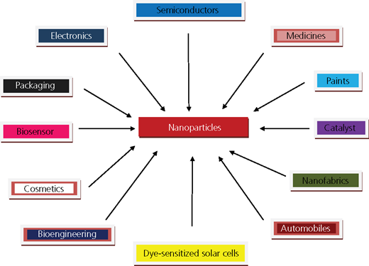
Metal NPs have been reported to have excellent chemical reactivity and potential applications in biosensing, drug delivery, detection of genetic disorders, gene therapy, catalysis, surface-enhanced Raman scattering, photography and DNA sequencing (Figure 1).5
NPs have unique properties compared to microparticles, with regard to shape, size and distribution.6 Silver (Ag)-based compounds have been used in medical applications since the nineteenth century.7 Silver nanoparticles (SNPs) have been used as antimicrobial agents in elevators and railway stations in China.8 Silver is usually used in the form of silver nitrate (AgNO3) to induce antimicrobial effects; when silver is used at the nanoscale, its enhanced surface area increases the magnitude of action toward microbes. SNPs can inhibit cell transduction, cell lysis and growth.9
Current studies have revealed that the use of SNPs in the treatment of wound healing causes reduction of local matrix metalloproteinase activity and enhances neutrophil apoptosis within the wounds.10 When SNPs were introduced in a rat model with burn injury, these caused reduction in the level of proinflammatory cytokines; SNPs play a vital role in the inhibition of the tumor necrosis factors alpha and activities of interferon gamma, which are responsible for inflammation.11,12
Nanosilver-based design for wound dressing was first discovered by Dr Robert Burrell in 1995. The name of the dressing was Acticoat; nowadays, it is being sold worldwide by Smith and Nephew.13 SNPs are coated with polymethyl methacrylate and used in bone cements as artificial joint replacements. When polypropylene mesh was coated over nanosilver particles, it showed tremendous antimicrobial activity.14
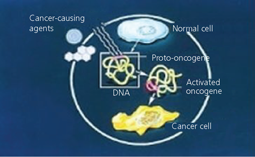
A proto-oncogene is a normal gene that regulates cell growth in a controlled fashion, but it can be converted into an oncogene by way of mutations and overexpression. Activated oncogenes are responsible for inducing tumor and cancer in healthy cells. Cancer is the fastest spreading and most fatal disease all over the world; malignant cells undergo rapid and uncontrolled cell division in the human body.15
Cancer cells proliferate through different steps of mutation, on which anticancer drugs exert their mechanisms of action (Figure 2). The philosophy of using anticancer drugs is to inhibit one or more actions/steps of mechanisms of cancer cell proliferation or inhibit an organelle to work for the cells.
2 Methods of NP fabrication
NPs are fabricated by physical, chemical and biological methods. SNPs (3–40 nm) are obtained by chemical reduction of metal slat of silver tetrafluoroborate (AgBF4) by sodium borohydride in water.16 Very expensive conventional methods have been used for the fabrication of NPs while showing hazardous effects (Figure 3).
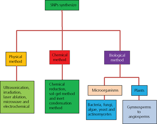
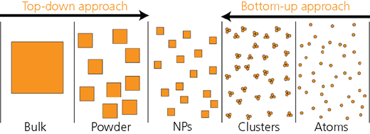
Thus, scientists have put their attention toward using green synthesis for the formation of NPs due to obtain their precise shape and controlled structures. Two approaches are preferred: top-down and bottom-up (Figure 4).17
Top-down approach: The lithographic technique is used to break down the bulk material particles into nanoscale size.18
Bottom-up approach: Atoms are self-assembled by electrochemical deposition and sonodecomposition to form new nuclei, which grow into NPs of desired sizes.
3 Green synthesis of nanosilver particles
3.1 Role of microorganisms in NP fabrication
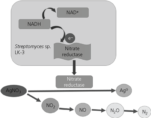
Amulyavichus et al.19 first reported that SNPs were isolated from silver mine using Pseudomonas stutzeri AG259. The mechanisms involved in the resistance of microorganisms are responsible for alteration of solubility and toxicity by way of reduction.20 In vitro synthesis by silver using bacteria (Bacillus licheniformis) causes secretion of alpha nicotinamide adenine dinucleotide phosphate (NADP) and NADPH enzymes, which are responsible for nitrate reductase, which efficiently converts silver ion into metallic silver (Figure 5).21
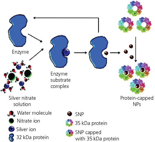
If the formations of SNPs by bacteria and fungi are compared, fungi show outstanding production of NPs compared with bacteria due to the ability of fungi to emit a greater amount of proteins that directly translates to enhance the production of NPs.22
3.2 Phytofabrication of SNPs
Plant extracts could be utilized as a source for the fabrication of SNPs; chemical constituents such as secondary metabolites present in plant extracts act as reducing and capping agents. Silver nitrate, aurum chloride and titanium chloride have been used for the formation NPs such as silver, gold and titanium by using plant extracts.25,26
Plants consist of a complicated set of antioxidant metabolites and enzymes that work against oxidative damage to cellular components. Phytochemicals such as polyphenols, ascorbic acid, triterpenes, alkaloids, alcoholic compounds, saponins, flavonoids, sterols, fructose, glucose, protein and β-phenylethylamines have been used for the reduction of silver ion into metallic silver.22,27–29
Different parts of plants contain a variety of functional groups of secondary metabolites that cause reduction of silver ions.30,31 After the formation of SNPs, characterization of NPs by using different surface techniques – for example, SEM – is then carried out to determine the morphology of NPs at submicron to micron scales.
Fourier-transform infrared spectroscopy can be used to identify functional groups attached to the surface of NPs, whereas X-ray powder diffraction is used to determine the crystal structure and phase identification of NPs.32,33 Metallic NPs have been synthesized by using different phytoextracts shown in Table 1.
Some phytofabricated nanoparticles
| Plant | Plant parts | Biomolecules involved | Type of NP | Size and shape | References |
|---|---|---|---|---|---|
| Acalypha indica | Leaf | Quercetin | Silver | 20–30 nm; spherical | Sun et al.34 |
| Achyranthus aspera | Leaf | Polyols | Silver | 20–30 nm; spherical | Krishnaraj et al.35 |
| Allium sativum | Leaf | Sucrose and fructose | Silver | 4–22 nm; spherical | Daniel et al.36 |
| Aloe vera | Leaf | Proteins | Silver, gold | 50–350 nm; spherical and triangular | Von White et al.37 |
| Azadirachta indica | Leaf | Salanin, nimbin, azadirone and azadirachtin | Silver, gold | 7·5–6·5 nm; spherical, triangular, quasi-spherical | Chandran et al.38 |
| Astragalus gummifer | Leaf | Proteins | Silver | 13·1±1·0 nm; spherical | Thirumurugan et al.39 |
| Andrograhis paniculata | Leaf | Hydroxyflavones; catechin | Silver | 28 nm; spherical | Kora and Arunachalam40 |
| Anacardium occidentale | Leaf | Polyols and proteins | Gold, silver, gold core–silver shell, gold–silver alloy | — | Jayashree and Vani41 |
| Allium cepa | Leaf | Vitamin C | Gold | Approximately 100 nm | Sheny et al.42 |
| Avena sativa | Leaf | Phenolics | Gold | 6–20 nm | Parida et al.43 |
| Argemone Mexicana | Leaf | Terpenoid | Silver | — | Shankar et al.44 |
| Boswellia ovalifoliolata | Leaf | Tannins and quinines | Silver | 30–40 nm | Singh et al.45 |
| Brassica juncea | Leaf | Oxalic acid | Gold | — | Ankanna et al.46 |
| Bacopa monniera | Leaf | Phenolics | Silver | 15–120 nm | Marshall et al.47 |
| Boswelliaovali foliolata | Bark and leaf | Polyols, heterocyclic components | Silver | — | Mahitha et al.48 |
| Bauhinia variegate | Leaf | Polyphenols or flavonoids | Gold | 43–145 nm | Jha et al.49 |
| Cinnamomum camphora | Leaf | Polyols, heterocyclic components | Gold and silver | 55–80 nm | Kumar and Yadav50 |
| Capsium annuum | Leaf | Proteins | Silver | 15–20 nm | Huang et al.51 |
| Cinnamomum zeylanicum | Leaf | Terpenoid | Silver | 50–100 nm | Li et al.25 |
| Carica papaya | Leaf | Hydroxyflavones and catechins | Silver | 15 nm | Sathishkumar et al.52 |
| Coriander | Leaf | Proteins and amino acids | Gold | 6·75–57·91 nm | Kora and Arunachalam40 |
| Cariandrum sativum | Leaf | Terpenoid, flavonoids | Silver | 26 nm | Narayanan and Sakthivel53 |
| Curcuma longa | Leaf | Flavones, alkaloid or proteins | Silver | — | Sathyavathi et al.54 |
| Catharanthus roseus | Leaf | Proteins and amino acids | Silver | 48–67 nm | Sathishkumar et al.55 |
| Camellia sinensis | Leaf | Polyphenolic compounds | Gold | 25 nm | Kannan et al.56 |
| Centella asiatica | Leaf | Terpenoid, flavonoids | Silver | — | Boruah et al.57 |
| Chenopodium album | Leaf | Oxalic acid | Silver, gold | 12, 10 nm | Palaniselvam et al.58 |
| Coleus aromaticus | Leaf | Flavonoids | Silver | 40–50 nm | Dwivedi and Gopal59 |
| Cycas circinalis | Leaf | Proteins and amino acids | Silver | 2–6 nm | Vanaja and Annadurai60 |
| Citrullus colocynthis | Leaf | Polyphenols with aromatic ring and bound amide region | Silver | 31 nm | Jha et al.49 |
| Chrysanthemum morifolium | Leaf | Flavonoids | Silver | 20–50 nm; spherical | Priya et al.61 |
| Chrysopogon zizanioides | Leaf | Terpenoid | Silver | 85–110 nm, cubic | He et al.62 |
| Chrysanthemum indicum | Leaf | Polyphenols or flavonoids | Silver | 37·71–71·99 nm; spherical | Arunachalam and Annamalai63 |
| Datura metel | Leaf | Plastohydroquinone or plastrocohydroquinol | Silver | 16–40 nm | Arokiyaraj et al.64 |
| Desmodium triforum | Leaf | Water soluble antioxidative silverent such as ascorbic acids | Silver | 5–20 nm | Kesharwani et al.65 |
| Dioscorea bulbifera | Leaf | Polyphenols or flavonoids | Silver | 8–20 nm Average 75 nm | Ahmad et al.66 |
| Diopyros kaki | Leaf | Terpenoid and reducing sugars | Platinum | 2–12 nm | Ahire et al.67 |
| Dioscorea oppositifolia | Leaf | Polyphenols with aromatic ring and bound amide region | Silver | 14 nm | Song et al.68 |
| Dalbergia sisso | Leaf | Polyphenols or flavonoids | Gold and silver | 5–80 nm and 5–55 nm | Maheswari et al.69 |
| Elettaria cardamomom Maton | Leaf | Alcohols, carboxylic, acids, ethers, esters and aliphatic amines | Silver | — | Singh et al.70 |
| Eclipta prostrate | Leaf | Flavones and proteins | Silver | 35–60 nm, triangles, pentagons, hexagons | Gnanajobitha et al.71 |
| Euphorbiaceae latex | Leaf | Flavonoids, proteins and terpenoids | Copper/silver | 18 nm silver and 10·5 nm copper | Rajakumar and Rahuman,72 Patil et al.73 |
| Emblica officinalis | Leaf | Polyphenols or flavonoids | Silver and gold | 10–20 nm and 15–25 nm | Valodkar et al.74 |
| Euphorbia hirta | Leaf | Flavonoids | Silver | — | Ankamwar et al.75 |
| Eucalyptus hybrid leaf | Leaf | Flavanoid and terpenoid | Silver | 50–150 nm; spherical | Elumalai et al.76 |
| Ficus benghalensis | Leaf | Polyphenols | Silver | 16 nm | Dubey et al.77 |
| Geranium | Leaf | Phenolics | Silver | 16–40 nm; quasilinear superstructure | Jegadeeswaran et al.78 |
| Gliricidia speium | Leaf | Polyphenols or flavonoids | Silver | 10–50 nm; spherical | Shankar et al.79 |
| Gardenia jasminoides Ellis | Leaf | Geniposide, chlorogenic acid, crocins and crocetin | Palladium | 3–5 nm | Raut et al.80 |
| Glycyrrhiza glabra | Leaf | Flavonoids, terpenoid, thiamine | Silver | 20 nm | Jia et al.81 |
| Hibiscus cannabinus | Leaf | Ascorbic acid | Silver | 9 nm | Dinesh et al.82 |
| Hydrilla verticilata | Leaf | Proteins | Silver | 65, 55 nm | Bindhu and Umadevi83 |
| Humulus lupulus | Leaf | Terpenoid | Gold | — | Sable et al.84 |
| Henna | Leaf | Alkaloids, flavonoids | Gold, silver | 21, 39 nm | Rai et al.85 |
| Hibiscus rosa-sinensis | Leaf | Polyphenols with aromatic ring and bound amide region | Silver | 4 nm; spherical | Kasthuri et al.86 |
| Ipomea carnea | Leaf | Polyphenols and hydroxyl groups | Silver | 30–130 nm | Philip87 |
| Jatropha curcas | Leaf | Curcacycline A (an octapeptide), curcacycline B (a non-apeptide), curcain (an enzyme) | Zinc sulfide, lead | 10 nm; 10–12·5 nm | Daniel et al.,88 Bar et al.89 |
| Justicia gendarussa | Leaf | Polyphenol and flavonoids | Gold | 27 nm | Hudlikar et al.90 |
| Lantana camara L. | Leaf | Carbohydrates, glycosides and flavonoids | Silver | 12·55 nm | Fazaludeen et al.91 |
| Leonuri herba L. | Leaf | Polyphenols and hydroxyl groups | Silver | 9·9–13·0 nm | Sivakumar et al.92 |
| Lxora coccinea | Leaf | Alcohols, carboxylic, acids, ethers, esters and aliphatic amines | Silver | 13–57 nm; spherical | Im et al.93 |
| Mentha pipertia | Leaf | Menthol | Silver, gold | 5–150 nm; spherical | Karuppiah and Rajmohan94 |
| Musa paradisiacal | Leaf | Proteins | Silver | 20 nm | MubarakAli et al.95 |
| Macrotyloma uniflorum | Leaf | Polyphenols with aromatic ring and bound amide region | Gold | 14–17 nm | Bankar et al.96 |
| Mirabilis jalapa | Leaf | Polyols | Gold | 100 nm | Aromal et al.97 |
| Morinda pubescens L. | Leaf | Hydroxyflavones, catechins | Silver | 25–50 nm | Vankar and Bajpai98 |
| Medicago sativa | Leaf | Alcohols, carboxylic, acids, ethers, esters and aliphatic amines | Silver | 2–20 nm | Jancy and Inbathamizh99 |
| Memecylonedule | Leaf | Water-soluble carbohydrates | Silver and gold | 5–55, 50–90 nm | MubarakAli et al.95 |
| Morinda citrifolia | Leaf | Phenolic compounds alcohols, phenols and proteins | Silver | 32–55 nm, oval | Parashar et al.100 |
| Nerium indicum | Leaf | Polyphenols and hydroxyl groups | Silver | — | Ankamwar et al.75 |
| Nicotiana tabaccum | Leaf | Proteins and amino acids | Silver | 8 nm | Suman et al.101 |
| Nelumbo nucifera | Leaf | Protein and enzymes | Silver | 25–80 nm; spherical triangular | Prasad et al.102 |
| Ocimum sanctum L. | Leaf | Phenolic and flavanoid compounds, proteins, ascorbic acid, gallic acid and terpenoids | Silver, gold, platinum | Approximately 10, 4–30, 23 nm | Santhoshkumar et al.,103 Ahmad et al.,104 Ramteke et al.105 |
| Parthenium hysterophorous | Leaf | Hydroxyflavones and catechin | Silver | 10 nm | Soundarrajan et al.106 |
| Pedilanthus tithymaloides | Leaf | Protein and enzymes | Silver | 15–30 nm | Ashok Kumar107 |
| Pipper betle L. | Leaf | Proteins | Silver | 3–37 nm | Abbasi108 |
| Piper nigrum L. | Leaf | Proteins | Silver | 5–50 nm | Koduru et al.109 |
| Plumeria rubra L. | Leaf | Proteins | Silver | 32–220 nm | Garg110 |
| Parthenium leaf | Leaf | Flavones and anthocyanins | Gold | 50 nm; face-centered cubic | Patil et al.111 |
| Pelargonium graveolens | Leaf | Phenolic and flavanoid compounds, proteins, ascorbic acid, gallic acid and terpenoids | Silver | 16–40 nm | Parashar et al.112 |
| Phyllanthus maderaspatensis | Leaf | Proteins, flavones and terpenoids | Silver | 30–130 nm | Shankar et al.113 |
| Rosa rugosa | Leaf | Amines, alcohols | Silver, gold | 30–60 nm silver; 50–250 nm gold | Annamalai et al.114 |
| Sesuvium portulacastrum L. | Leaf | Proteins, flavones and terpenoids | Silver | 5–20 nm | Dubey et al.115 |
| Solanum xanthocarpum L. | Leaf | Phenolics, alkaloids and sugar | Silver | 10 nm | Nabikhan et al.116 |
| Sorghum bicolor Moench | Leaf | Polyphenols | Silver, iron | 10 nm, 50 nm | Amin et al.117 |
| Soybean | Leaf | Proteins and amino acids | Lead | Approximately 15 nm | Njagi et al.118 |
| Swietenia mahogani | Leaf | Polyhydroxy limonoids | Silver, gold and bimetallic alloy gold–silver | — | Petla et al.119 |
| Syzgium aromaticum | Leaf | Flavonoids | Gold | 5–100 nm | Mondal et al.120 |
| Sesbania drummondii | Leaf | Proteins and amino acids | Gold | 6–20 nm | Raghunandan et al.121 |
| Syzgium cumini | Leaf | Flavonoids | Silver | 100–160 nm | Nabikhan et al.116 |
| Syzgium cumini | Bark | Proteins, flavones and terpenoids | Silver | 20–60 nm | Prasad and Swamy122 |
| Santalum album | Leaf | Hydroxyflavones and catechins | Silver | 80–200 nm | Prasad and Swamy123 |
| Sorghum bicolor | Leaf | Hydroxyflavones | Silver and iron | — | Swamy and Prasad124 |
| Salanum torvum | Leaf | Phenolic and flavanoid compounds, proteins, ascorbic acid, gallic acid and terpenoids | Silver | 5–50 nm; spherical | Njagi et al.118 |
| Saraca asoca | Leaf | Hydroxyflavones and catechins | Silver | 87–102 nm, irregular | Ramamurthy et al.125 |
| Terminalia catappa L. | Leaf | Hydrolyzable tannins | Gold | 10–35 nm | Suman et al.101 |
| Trianthema decandra | Leaf | Hydroxyflavones and catechins | Silver | 10–50 nm | Ankamwar126 |
| Tridax procumbens L. | Leaf | Water-soluble carbohydrates | Copper peroxide | — | Geethalakshmi and Sarada127 |
| Tanacetum vulgare | Leaf | Hydrolysable tannins | Silver, gold | 16 nm silver, 11 nm gold | Annamalai et al.114 |
| Trapa bispinosa | Leaf | Silver | — | Gopalakrishnan et al.128 | |
| Tamarindus indica | Leaf | Protein and enzymes | Gold | 20–40 nm | Pandey et al.129 |
| Vitus vinifera L. | Leaf | Flavones and anthocyanins | Lead | 661 nm | Pavani et al.130 |
| Vitex negundo | Leaf | Flavonoids | Gold | 10–30 nm; face-centered cubic | Zargar et al.131 |
| Zingiber officinale | Leaf | Alkanoids, flavonoids | Silver, gold | 10 nm | Singh et al.132 |
4 Mechanism of action of SNPs toward microbes
Silver metal has been used in people’s daily life for different purposes such as jewelry, ornamentation and fine cutlery. Silver is the only metal which has the potential to resist microbes, dating back to the Phoenicians. Silver is used as an antimicrobial agent to fight against many microorganisms, numbering approximately 650 from diverse modules such as gram-negative and gram-positive bacteria, fungi and viruses.
In 1884, aqueous silver nitrate was used in the form of eye drops for newborn babies to prevent the transmission of Neisseria gonorrhoeae from infected mothers. Metals have many properties; silver is the metal that shows the least toxic effect in in vivo and in vitro.46
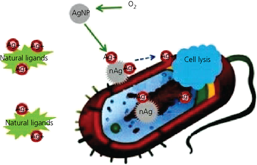
Silver is usually used in the form of salt, mostly silver nitrate, to show antimicrobial effect, but when SNPs are used, their physical parameters eventually changed, such as when huge surface areas have been exposed for the microbes. Biogenic SNPs have been used for evaluating their antimicrobial activities against different microbes. The antimicrobial properties of biofabricated SNPs are based on size, environmental conditions and capping agent. SNPs have a significant action against microbes; they have the ability to anchor to a bacterial cell and penetrate it (Figure 7). Silver is inert, but when it is in contact with moisture, it releases silver ions, so a positive charge indicating silver ions is reported to be vital for antimicrobial activities.133
Silver ions are able to form complexes with nucleic acids and preferentially interact with the nucleosides rather than with the phosphate groups of nucleic acids. Silver ions are obtained from silver or silver-containing compounds with notable antimicrobial properties; these silver ions may be incorporated into the substance and released slowly with time.134 Researchers have reported that a suitable bactericidal agent is formed due to a strong electrostatic force of attraction between positively charged SNPs and negatively charged bacterial cells.132,135,136 Two mechanisms have been reported for SNP resistance toward microbes.
4.1 First mechanism
SNPs can easily permeate into bacterial cell walls and cause cell death by changing the structure of bacteria; thus, pits that have formed on the cell surface of bacteria provide a way for NPs to accumulate.134 Ankanna et al. 46 reported that SNPs have been shown to accumulate inside the membrane and can subsequently penetrate into the cells then causing injury to the cell wall or cell membranes.
The exact mode of action of SNPs is that they bind to thiol groups (SH) of enzymes and then form the stable S–Ag bonds with thiol-containing compounds and are responsible for the deactivation of enzymes such as transmembrane energy generation and ion transport in the cell membrane.
When silver ions enter cells, these intercalate between the purine and pyrimidine base pairs, disrupting the hydrogen bonds between the two antiparallel strands and denaturing the DNA molecule. Bacterial cell lysis could be one of the reasons for the antibacterial property of silver NPs. NPs modulate the phosphotyrosine profile of bacterial peptides, in turn affecting signal transduction and inhibiting the growth of microorganisms.
Escherichia coli cells have been treated with SNPs, and SNPs have been found to be accumulated in the bacterial membrane, which results in an increase in the permeability of the cell wall and death of the cell. Gram-positive bacteria are less susceptible to silver than gram-negative bacteria. The reason is that the gram-positive bacterial cell wall has more peptidoglycan molecules than the gram-negative bacterial cell wall.
As the cell wall of gram-positive bacteria is thicker and peptidoglycans are negatively charged and silver ions are positively charged, more SNPs may get stuck to the peptidoglycans in gram-positive bacteria than in gram-negative bacteria. The decreased liability of gram-positive bacteria can also be simply explained by the fact that the cell wall of gram-positive bacteria is thicker than that of gram-negative bacteria.131
4.2 Second mechanism
It is proved by literature that DNA has the ability to destroy the cell membrane through an electron-release mechanism or free radical production then formation of SNPs playing a key role to make the membrane too much porous and accumulate to envelope protein precursor or destabilization of outer membrane, which finally leads to ATP leaking, which can eventually lead to cell death.46,137,138
The spectroscopic technique (electron spin resonance) has been recommended to analyze the formation of free radicals by SNPs.139,140 When bacterial cells come in contact with silver salts, the salts interact with thiol groups of many vital enzymes that exist in the cell membrane of bacteria which take up the silver ions, resulting in the inactivation of various functions in the cell and permanent injury to the cells.
Reactive oxygen species (ROS) are generated by silver ion to inhibit the respiratory enzyme of bacterial cell and attack the cell itself. Silver has a tendency to behave as an acid to react with the base present in the DNA of a cell – that is, sulfur (S) and phosphorus retarded the function of DNA ultimately causing cell death.141,142 It is reported that phosphorylation of protein substrates in bacteria causes influences in signal transduction of bacteria and inhibition of growth.143
Silver has a potential as antiseptic and disinfectant; it has the ability to interact with disulfide bonds of the glycoprotein/protein contents of microorganisms such as viruses, bacteria and fungi. SNPs and silver ions are responsible for changing the three-dimensional structure of proteins by interfering with disulfide bonds and block the functional operations of the microorganism.144–146
Biogenically fabricated NPs are gaining importance over chemical and physical methods because it is cost-effective and eco-friendly and can be easily scaled up for large-scale synthesis, and there is no need to use high energy, pressure, temperature and toxic chemicals.147–150
5 Mechanism of action of SNPs toward oncogenes
Cancer is a disease that is caused by the changes in genes that control the way cells function, particularly how they grow and divide. Genetic changes arise as the result of errors that occur in cell division because of DNA damage. In general, cancer cells have more genetic changes such as mutations in DNA than normal cells.
Akhtar et al.30 reported mechanisms in cancerous cell by using biomimetic NPs as novel controlling agents. Biogenic nanosilver has immense potential toward various cancer cell lines such as Hep 2, HCT 116, MCF-7, A549, HT-29 and Caco-2 (Figure 8).151
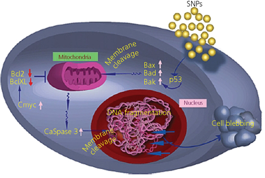
NPs, once attached to the cell membrane, damage the integrity of the membrane and trigger the activation of p53 protein. In turn, p53, a known activator of proapoptotic gene, activates Bax, Bad and Bak. These activated proteins are known to cause mitochondrial membrane leakage and release cytochrome complex, which, in a cascade reaction, activates caspase-3. Bcl-xL (B-cell lymphoma-extra large) and Bcl 2 (B-cell lymphoma 2) are proteins in humans encoded by the Bax gene Bcl-xL, corroborating manifestation of apoptosis and consequent cell blebbing. Phytofabricated SNPs possess great selectivity to cancer cells and can display potential application in cancer chemoprevention and chemotherapy. SNPs have striking antiangiogenic properties and the ability to block the activity of abnormally expressed signaling proteins.
5.1 In vivo mechanism of SNPs on cancerous cells
When SNPs are exposed to certain mammalian cells, some proteins such as interleukins, macrophage colony-stimulating factor and macrophage inflammatory proteins are activated and upregulated. SNPs can stimulate inflammatory cells. They play a vital role in the formation of ROS and damage the DNA of cancer cells; this ROS depletes glutathione in rat liver cell by way of damaging the mitochondria.
SNPs have large surface area-to-mass ratio, so they can provide a large functional surface to bind, absorb and carry other compounds such as drugs, probes and protein – for example, ligands have ability to find the receptor of targeted cells. Tumor cells are usually considered more acidic in nature, so SNPs release more in tumor cells than in normal cells. Currently, an in vivo study has examined the NP distribution with diameter greater than 100–150 nm to accumulate in tumors because of their higher extravasation in comparison with normal vasculature.152–154
The cytostatic effect of metallic silver on cancer cells is due to active physichemical interaction of SNPs with bimolecular active functional groups of intercellular protein and DNA. SNP administration caused significant reduction of proliferation in glioblastoma multiforme (GBM) cells.155,156 Both proliferative and mitotic indices were significantly lower in the SNP group in comparison with control and placebo groups. Biogenic SNP accumulate in the nucleus of GBM cells, where they cause chromosome instability, malignancy in tumor cells and mitotic arrest.157
Medicated SNPs bind with functional groups to inhibit the overexpression of cell division and signaling cascade; SNPs have the potential ability to cure GBM against cancer. Tumor-bearing mice have a longer survival time when SNPs are injected; the effect of SNPs is more potent than that of vincristine treatment.
The SNP active surface can directly induce the generation of free radicals; it causes DNA damage and disruption of mitochondrial membrane, potentially leading to releases of cytochrome complex and mitochondria-dependent apoptosis. The majority of anticancer drugs initiate apoptosis causing caspase IX overexpression, which, after activation, leads to apoptosis.
6 Conclusion
Cancer is treated by using different conventional treatments such as surgery, radiotherapy and chemotherapy, which pose harmful effects on healthy tissues of the body. Biogenically fabricated SNPs have increased their importance due to its eco-friendliness. Green synthesis is now overcoming the limitations of physical and chemical methods of formations of metallic NPs. Phytofabricated nanosilver particles are less time-consuming, cost-effective and non-toxic. Silver metal has shown significant potential as an antimicrobial and anticancer agent. When silver is used as phytofabricated SNPs with increased surface area, it has enhanced efficiency against microbes and oncogenes. This current review has summarized the mechanisms of action of SNPs in antimicrobial and anticancer activities.
