

Laser surface texturing (LST) has become a pivotal technique in biomedical surface engineering, providing unparalleled precision in modifying material surfaces to enhance implant performance. This review integrates recent advancements in femtosecond and ultrafast laser technologies, offering novel insights into how these methods enhance osseointegration, antibacterial properties, and tissue regeneration in biomedical implants. Unlike previous reviews, this paper delves into specific laser parameters and their influence on surface morphology, wettability, and biocompatibility, particularly in orthopaedic and dental applications. A detailed comparison of LST techniques highlights their role in reducing implant failure due to bacterial contamination and inadequate tissue integration. The scalability of LST for clinical and commercial applications is also explored, emphasizing its potential as a transformative tool for improving implant longevity and reducing post-surgical complications. Furthermore, this review critically evaluates challenges and limitations, including scalability issues, standardization concerns, and long-term clinical validation. By synthesizing the latest advancements, this work provides a comprehensive foundation for future developments in laser-assisted biomedical surface engineering, positioning LST as a prominent approach in next-generation biomedical materials and medical device innovation.
1. Introduction
The process of surface texturing involves the formation of a unique design or texture on a given work surface. This methodology offers a distinct advantage in altering the surface morphology to augment the tribological characteristics of the material, such as its load-carrying capacity, durability against abrasion, and coefficient of friction.1–4 A variety of techniques, including chemical vapour deposition (CVD), sandblasting, electric discharge texturing, hot embossing, lithography, and laser texturing, are utilised to improve tribological properties through the application of coatings and textures. Among all techniques, laser surface texturing (LST) has gained popularity among researchers and industrialists as a viable texturing technique because of its exceptional precision, high efficiency, environmental friendliness, and capacity to customise surface properties at the micro- and nanoscale levels. During laser texturing, a high-intensity laser beam impacts the surface of the workpiece, causing ablation and resulting in both material melting and vapourisation. Laser texturing has been efficaciously implemented across various engineering endeavours, including coating, tribological, and biomedical ones. LST has created a variety of patterns and textures to improve the material’s tribological performance. Moreover, the treatment enhances the material’s durability and coefficient of friction. Arthritis is considered one of the most severe conditions globally. In India, a staggering 180 million individuals, constituting approximately 15% of the population, are affected by arthritis-related conditions. This issue is becoming more serious due to the increase in older individuals, obese people, and diabetes sufferers.5 Although there have been significant advancements in orthopaedic implant techniques, only a minor proportion of knee and hip implants endure beyond 20 years, with just 90% lasting between 10 and 15 years.6–10
The orthopaedic industry is poised to expand significantly, with projections indicating a rise from a total of USD 105.18 billion in 2019; the industry is projected to reach an impressive sum of USD 206.64 billion by the year 2024, fuelled by escalating global demand.11–14 With its exceptional precision and ability to tailor surface characteristics at the micro- and nanoscale levels, laser texturing offers unique advantages for enhancing the performance of biomedical materials, medical devices, and tissue-engineered constructs. The present review delves into the cutting-edge implementations, tenets, and progressions of laser texturisation within biomedical engineering. Our emphasis lies particularly on its capacity to transform the milieu of regenerative medicine and medical apparatus innovation.
This review provides a comprehensive analysis of recent advancements in LST for biomedical applications, focusing on orthopaedic and dental implants. We explore the latest innovations in femtosecond and ultrafast laser technologies, highlighting their impact on improving implant biocompatibility, antibacterial properties, and osseointegration. This review critically evaluates the role of specific laser parameters in enhancing surface morphology and tissue integration, comparing various LST techniques and their biomedical relevance. In addition, we discuss the challenges, limitations, and future research directions for LST in biomedical engineering, offering insights into its potential for commercial scalability. Synthesising the most recent studies provides a foundation for future advancements in laser-assisted biomedical surface engineering.
2. Methodology
Studies were selected based on their relevance to biomedical applications, specifically on laser techniques that enhance biocompatibility, antibacterial properties, and tissue integration. Articles primarily discussing orthopaedic and dental implants were prioritised, while studies unrelated to medical applications were excluded.
The selected literature was then analysed and categorised according to the type of laser technology, the biomedical application, and the materials used in the implants (e.g. titanium alloys, zirconia, polymers). Special emphasis was placed on recent advances in femtosecond and ultrafast laser technologies and how they contribute to improved implant performance. The review also critically compares different laser texturing techniques and their impact on cellular responses, implant longevity, and antibacterial properties.
This structured approach enabled a comprehensive evaluation of the advancements in LST for biomedical applications while also identifying gaps in the current literature and suggesting future research directions.
3. Types of laser processing
From the literature survey, various types of laser texturing techniques used to generate texture on the materials were found. The detailed discussion about types of laser processing techniques used in laser texturing is as discussed as follows.
3.1 Direct laser ablation
Through the utilisation of a laser beam, solid surfaces undergo irradiation, resulting in the removal of materials from said surface by way of the process known as laser ablation. At heightened levels of laser flux, the interplay between the laser beam and matter triggers surface material vaporisation. The evaporated materials can subsequently undergo ionisation to form plasma. The selective elimination of material by way of laser ablation yields a distinct surface morphology replete with singular design motifs such as microtextures and ridges.15–17
Laser texturing through laser ablation is effective and used in various engineering and bioengineering applications. Fiorucci et al.18 fabricated a groove pattern with 30 µm width and 10 μm depth on titanium alloy Ti-6Al-4V through nanosecond laser ablation, which is suitable for biomedical applications. Using femtosecond direct laser ablation, Wang et al.19 observed a cone array–like microstructure on the surface of monocrystalline silicon with spike depths of up to 8 μm. Melo-Fonseca et al.13 fabricated microscale pillars with a pyramid geometry on Ti-6Al-4V titanium alloy utilising Nd:YAG laser ablation processing to achieve a textured surface. As depicted in Figure 1, the laser ablation process creates a well-defined groove pattern, enhancing the ceramic surface’s mechanical properties and tribological performance. The precise control over groove dimensions is critical in biomedical applications where surface features can directly impact cell adhesion and tissue integration.20 Laser processing was executed utilising Nd:YAG laser, featuring a 10 ns pulse width and a wavelength of 1064 nm. The computerised system utilised a software interface to regulate the laser power. Initially, the beam underwent a perpendicular bending process by way of a scanner before being concentrated through a focusing lens. Afterwards, green ZrCb ceramics were placed on the laser system’s three-dimensional XYZ stages, which possess an exceptional precision of 10 pm and are utilised for laser texturing applications. The computer that interfaces with this laser system is responsible for generating the intricate laser texture pattern.
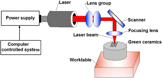
A visual representation of laser-induced surface texturing on green ceramic material is presented. Adapted from Liu et al.20
A visual representation of laser-induced surface texturing on green ceramic material is presented. Adapted from Liu et al.20
3.2 Direct laser interference patterning
One notable methodology for LST is laser interference, which is called direct laser interference patterning (DLIP). This technique generates a cyclic arrangement of characteristics by utilising coherent laser beam interference. The outcome manifests as localised heating of the work surface that occurs periodically due to photo-thermal interaction between the laser and the work surface.21,22 The migration of materials from regions of maximum interference to those of minimum is attributed to the surface tension gradient that arises due to a temperature gradient.23 DLIP can produce periodic surface geometries, such as line, dot, and cross-like patterns, on diverse material surfaces.24 DLIP coatings are currently involved in advancing biophotonic sensing cells (BICELLs) for optical biosensor development.25 The researchers’ findings evince that ultrashort pulsed direct laser-induced periodic surface structures (LIPSSs) is a valuable technique for generating recurrent surface patterns on steel surfaces and is aptly suited to uphold and bolster bacterial growth.26
A periodic variation in the form of a line is achieved by implementing a two-beam setup, as shown in Figures 2(a) and 2(b). Figure 2 demonstrates how DLIP can generate intricate surface topographies essential for biomedical implants. These periodic patterns, such as lines and dots, enhance biocompatibility by promoting osseointegration and antibacterial properties, making DLIP a promising technique in regenerative medicine. This phenomenon is explained in detail in Equation 1.28
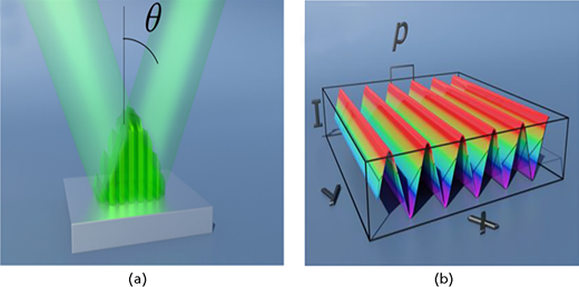
The system of interference between two laser beams, resulting in precise surface patterns such as lines, dots, and cross-like structures (a).27 These periodic surface textures are generated by controlling the number and angle of beams (b).27 Such patterns are critical in biomedical applications, as they enhance cell adhesion, promote osseointegration, and can be tailored to exhibit antibacterial properties on implant surfaces. These patterns’ spatial period (P) directly influences cell behaviour and tissue regeneration, making this technique valuable in improving implant performance27
The system of interference between two laser beams, resulting in precise surface patterns such as lines, dots, and cross-like structures (a).27 These periodic surface textures are generated by controlling the number and angle of beams (b).27 Such patterns are critical in biomedical applications, as they enhance cell adhesion, promote osseointegration, and can be tailored to exhibit antibacterial properties on implant surfaces. These patterns’ spatial period (P) directly influences cell behaviour and tissue regeneration, making this technique valuable in improving implant performance27
It is feasible to fabricate diverse patterns of geometries, incorporating structures resembling dots, pillars, and lamellae, by adjusting the number of beams, intensities, and polarisation. DLIP enables a rapid and seamless approach to surface patterning across multiple materials. The distance between beams impacts the arrangement of texture characteristics produced by laser interference, the separation between the laser and target, and the wavelength of the laser.29,30
Several engineering components and biomaterials have also been subjected to LST using laser interference. The researcher employed the direct laser interference technique to fabricate periodic micropatterns, in both one and two dimensions, on surfaces made of 100Cr steel.23 Guenther and colleagues31 utilised the DLIP technique on polymeric substrates, particularly polyimide and polystyrene, to showcase how surface topography impacts bacterial adherence patterns both in vivo and in vitro. The findings disclosed that bacteria tend to adhere to linear, one-dimensional formations. Conversely, complex three-dimensional arrangements resembling lamellae impede the development of biofilm. Nanoscale designs on silicon substrates have also been created using laser interference lithography. By increasing the power to 0.600 mW/cm2, studies have revealed that a 20 mm (W) by 20 mm (L) region can attain uniform dot arrangements, with a half-pitch measurement approaching 190, 250, and 370 nm.32 A team of researchers further establishes the efficacy of ultrashort pulsed DLIP as a technique for generating repetitive surface structures on steel material, which is conducive to both retaining and augmenting bacterial growth. Morales et al.33 investigated that Ti-6Al-4V’s texture homogeneity would be affected by laser parameters during DLIP, and the study team discovered an 80%–90% improvement in texture homogeneity.
3.3 Laser surface texturing by way of laser shock processing
Recently, sophisticated LST methodologies grounded on laser shock processing (LSP) have surfaced to tackle the impediments of laser ablation and interference. LSP embodies a state-of-the-art and refined surface engineering methodology that utilises shock waves generated by lasers to instigate compressive residual stress and fortifying impacts on the designated surface.34,35 The generation of surface texture transpires through plastic deformation induced by laser shock without any accompanying heat effect.36 Figure 3(a) illustrates a diagrammatical depiction of laser surface treatment utilising laser-induced plasma (LIP). A non-translucent film is applied onto the intended material to absorb the energy from the laser. A transparent enclosure encompasses the coating. The LIP technique induces plasma through interaction between the laser pulse and opaque coating instead of its targeted material.37
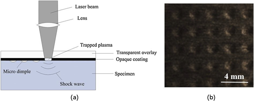
(a) Diagram illustrating the LSP process and (b) surface topography characterised by dimpled features generated by way of LSP.37
(a) Diagram illustrating the LSP process and (b) surface topography characterised by dimpled features generated by way of LSP.37
The plasma expansion is confined by a transparent enclosure, leading to the generation of a laser-induced shockwave that propagates within the target material with an immense peak pressure measured in giga-pascals. Once this shockwave surpasses the dynamic yield strength of the target materials, ultrahigh strain rate plastic deformation ranging from 105/s to 106/s takes place on the surface of processed samples.38 As a result, LSP can produce a surface texture that includes micro- or mini-dimples as depicted in Figure 3(b). Numerous studies have demonstrated that LSP can significantly improve the longevity of metallic materials.39–41 The increased resistance to wear is mainly attributed to the synergistic impact of surface hardening and residual stress.41 Li et al.37 produced microdimple arrays on copper through the employment of LSP methodology. The processed surfaces demonstrated superior tribological performance, characterised by diminished abrasive and adhesive wear compared with the untextured surface. Lim et al.42 showed a significant increase in microhardness values of duplex stainless steels from 250 HV to 310 HV following LSP treatment, resulting in a commendable reduction in wear rate by approximately 16%. Nevertheless, the low efficiency of the LSP process due to the need for individual fabrication of microfeatures such as dimples (as shown in Figure 3(b)) limits its scalability. It hinders practical applications within natural industries despite its ability to generate surface-hardening effects for improved wear resistance. In conclusion, Table 1 illustrates the unique advantages of each LST technique based on its specific application. For example, laser ablation is highly versatile, allowing for flexibility with different materials, whereas DLIP is particularly effective in producing nanoscale patterns that suit biomedical implants. This comparative analysis emphasises the critical need to choose the proper method to enhance biocompatibility and mechanical properties performance.
Comparison of laser surface texturing (LST) techniques (the checkmark symbol ✓ indicates the level. A rating of ✓✓✓✓✓ denotes an extremely high level, while a single checkmark denotes a very low level)
| Criteria | Laser ablation | Laser interference | Laser shock processing |
|---|---|---|---|
| Flexibility | ✓✓✓✓ | ✓✓✓✓ | ✓✓✓✓ |
| Efficiency | ✓✓✓ | ✓✓✓✓✓ | ✓✓✓ |
| Texture feature resolution limit | 1 μm for titanium43 | 0.1 μm for silicon substrate32 | 10 μm for aluminium alloy44 |
| Surface hardening effect mechanism | Heat-induced phase transformation (only for some specific metals) | Heat-induced phase transformation (only for some specific metals) | Surface plastic deformation for all metallic materials |
| Applicable materials | Metals, polymer, ceramics, and composite materials | Metals, polymer, ceramics, and composite materials | Only metals |
3.4 Laser parameter’s effects on surface texturing
The modulation of laser processing parameters, including laser power intensity, spot size, scan velocity, and pulse repetition frequency, can efficiently regulate LST procedures’ surface morphology and feature dimensions. Furthermore, the choice of laser type can also play a role in regulating these aspects. Comprehending the influence of surface texturing is paramount in refining LST methodologies and enhancing the tribological efficacy of materials post-treatment. The laser parameters are pivotal in ascertaining the efficiency and outcome of LST. The potency of the laser has a considerable influence on the dimensions and profundity of texture characteristics in ablation-based LST. Grabowski et al.45 conducted a comprehensive investigation into the impact of laser power intensity on surface morphology following laser processing of 316L steels, Ti-6Al-4V alloy, and AlSi7 alloy. The findings indicated a nearly linear progression in ablation depth across all three metallic materials as the laser power intensity increased. The magnitude of plastic deformation that occurs during laser processing, specifically in LSP-based LST, is contingent on the intensity of laser power. The shockwave pressure is crucial in moulding the surface morphology and enhancing the hardness of treated specimens.46 Zhou et al.47 generated microgrooves by way of a picosecond laser, varying the laser power, repetition frequencies, scan speeds, and scan numbers. The research findings demonstrate that the calibre of groove ablation is significantly influenced by laser power and scan speed. To achieve an optimal grade groove microstructure, it is recommended to utilise a laser with a 20 W output power and repetition rate set at 400 kHz and maintain a scan speed within the range of 1000–2000 mm/s. This approach was detailed in the previous analysis. Figure 4 clearly shows the distinct surface textures produced by different laser parameters. The smoother textures, as seen in the SEM images, facilitate better cell attachment, while the rougher, more jagged textures improve osseointegration by mimicking the structure of natural bone. As shown in Figure 4(a), the ablated surface manifests a ragged appearance with dispersed residual fragments embedded within the groove and a disrupted edge surrounding it. Although appearing inverted, Figure 4(b) still harbours substantial residue within the groove. In contrast to the grooves depicted in Figures 4(a) and 4(b), those shown in Figures 4(c) and 4(d) boast exceptional quality, featuring smooth bottoms and sharp edges. Despite this, a region remains affected by heat and sporadic bursts of debris and metal. The area impacted by thermal effects has been significantly reduced, as expected, and the landscape displays noticeable enhancements when contrasted with Figures 4(a) and 4(b). Moreover, the ridges display unique LIPSSs that arise from the interplay of incident laser radiation and surface plasma polarisation on the titanium alloy’s surface under specific conditions. This is exemplified by the partially magnified SEM images positioned to the right of Figure 4.
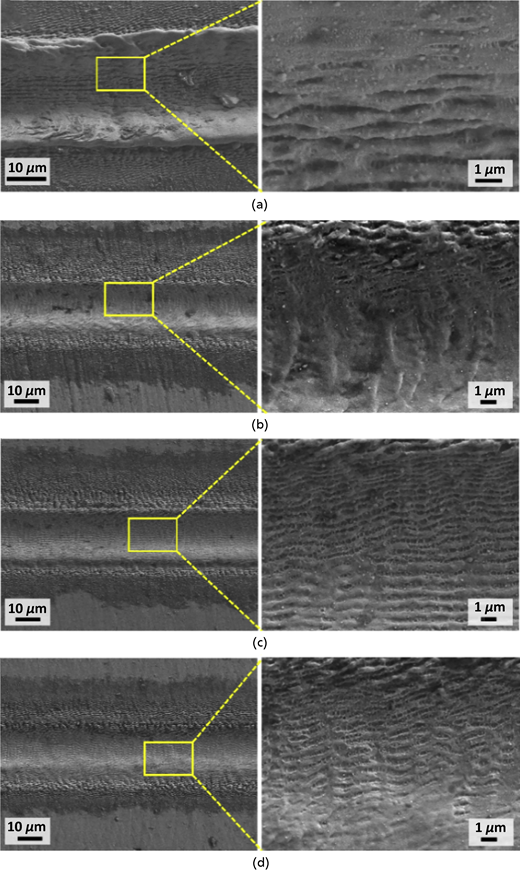
SEM images of the titanium alloy surface formed under picosecond laser treatment. (a) p = 25 W, V = 500 mm/s, n = 5, f = 400 kHz, (b) p = 25 W, V = 1000 mm/s, n = 10, f = 100 kHz, (c) p = 30 W, V = 2000 mm/s, n = 10, f = 400 kHz, and (d) p = 20 W, V = 2000 mm/s, n = 10, f = 400 kHz47
SEM images of the titanium alloy surface formed under picosecond laser treatment. (a) p = 25 W, V = 500 mm/s, n = 5, f = 400 kHz, (b) p = 25 W, V = 1000 mm/s, n = 10, f = 100 kHz, (c) p = 30 W, V = 2000 mm/s, n = 10, f = 400 kHz, and (d) p = 20 W, V = 2000 mm/s, n = 10, f = 400 kHz47
4. Type of lasers
Commercial pulse lasers can be classified into four categories depending on the duration of their laser pulses: millisecond, nanosecond, picosecond, and femtosecond. The length of the laser pulse plays a crucial role in numerous factors associated with LST processes, such as heat input during laser ablation and interference, along with the rate of plastic deformation in LSP. These variables ultimately determine the efficacy and efficiency of the overall process.48–51 Bathe et al.51 generated surface patterns on a grey cast iron utilising three distinct lasers. Figure 5 shows the textured surfaces modified by way of millisecond, nanosecond, and femtosecond lasers. Due to the high energy and prolonged pulse duration of the millisecond laser, it is evident that the treated surface experienced partial melting, resulting in the expulsion of molten components from the dimple. The discharged elements coalesced to form a bulge around the indentation. In contrast to samples treated with millisecond and nanosecond lasers, the sample treated with femtosecond laser exhibited the most seamless surface and precise dimple profile. Ahuir-Torres et al.52 utilised nano- and picosecond lasers to conduct a comparative assessment of the surface morphologies of Ti-6Al-4V alloys following ablation-based laser shock treatment. The research findings indicate that the shift from a nanosecond laser to a picosecond laser led to a noteworthy decrease in the extent of the heat-affected region. In laser ablation, a brief pulse duration can reduce the unwanted thermal impact and improve the precision of microstructuring. Bhaduri et al.1 examined and contrasted the surface topography of planar tungsten carbide blocks after femtosecond and nanosecond ablation during LST. The textures created using femtosecond lasers included a few nanoripples, but samples treated with nanosecond lasers lacked this feature.
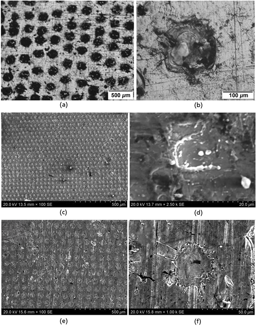
The effects of different laser types on the surface morphologies of grey cast iron during ablation-based laser surface texturing (LST) are investigated in this study. Specifically, we consider three distinct lasers with varying pulse durations: (a) millisecond laser, (c) nanosecond laser, and (e) femtosecond laser. The corresponding magnified regions for each indentation in images (a), (c), and (e) are denoted as (b), (d), and (f).51
The effects of different laser types on the surface morphologies of grey cast iron during ablation-based laser surface texturing (LST) are investigated in this study. Specifically, we consider three distinct lasers with varying pulse durations: (a) millisecond laser, (c) nanosecond laser, and (e) femtosecond laser. The corresponding magnified regions for each indentation in images (a), (c), and (e) are denoted as (b), (d), and (f).51
Recent studies have shown that femtosecond laser texturing offers unprecedented control over surface morphology, allowing for the precise creation of patterns that significantly improve biocompatibility and antibacterial properties. Wang et al. demonstrated the use of femtosecond lasers to fabricate microgrooves on titanium implants, enhancing cell proliferation and adhesion, which is critical for long-term osseointegration.
5. Surface texturing of biomaterials
The external layers of voluminous implants are primarily affected by biotic and abiotic factors. Augmenting surface characteristics constitutes the remedy for mitigating implant malfunction rates. Surface biomaterial modification is done to improve the mechanical, chemical, physical, and biocompatible qualities. According to a bone retrieval analysis, the average bone-to-implant contact for current orthopaedic implants is between 70% and 80%. As per the 2016–2017 Canadian Joint Registry Report, revision surgeries for hip and knee replacements constitute a significant proportion of orthopaedic procedures performed, accounting for 8.5% and 6.8%, respectively. These revisions are primarily necessitated by infections and inadequate osseointegration, which are the main culprits behind implant failure53 (Figure 6).
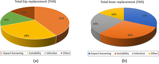
Boosting cellular proliferation by 15%–30% by way of exceptional osseointegration can reduce infections by an astounding 60%–90%.54 As a result, there will be lots of room for improved surface modification to enhance the performance of biomaterials. Surface concave and convex structuring are the two main divisions of surface modification. Surface concave structuring uses electrochemical, chemical, and mechanical processes for material removal. Sandblasting, shot peening, and laser peening are mechanical methods utilised to induce residual compressive surfaces for alteration. The efficacy of these techniques varies depending on factors such as media size, coverage, arc height, and other variables. Contrarily, surface convex texturing encompasses the application of materials by way of physical or chemical means. Many techniques, such as CVD, physical vapour deposition, solid-state diffusion bonding, and plasma spraying, are employed to fabricate curved surfaces that ultimately alter the density and porosity of bioimplant surfaces. More contact surface area is provided by these porous surfaces, which improves cell anchoring and proliferation. However, due to their several limitations such as contamination, non-uniformity, microcracks, and so on, they are not preferable for long-time run.55
6. Laser texturing in biomedical
The domain of inquiry that has undergone significant expansion in recent times pertains to the compatibility of a substance with host tissues. Materials exhibiting exceptional biocompatibility are deemed appropriate for employment in bone and tissue transplantation procedures.56 The development of biomaterial throughout the years demonstrates its widespread application in implants. Biomaterials commonly comprise metals, polymers, ceramics, and composites.57 The incidence of implant failure in subsequent surgeries and revision procedures is comparably elevated. The primary causal factors for such failures are inadequate osseointegration and bacterial infections. Present surface coating and modification methods are ineffective in ensuring enduring stability. Surface modification techniques such as CVD, grit blasting, acid etching, and anodic oxidation are quick and easy to employ, but their reproducibility is mediocre. The most auspicious solution in this circumstance is LST, as it can create surfaces that are biocompatible, antimicrobial, and favourable for prompt bone regeneration. LST exerts exceptional control over the process thanks to its meticulous manipulation of surface topography, morphology, wettability, and chemistry.
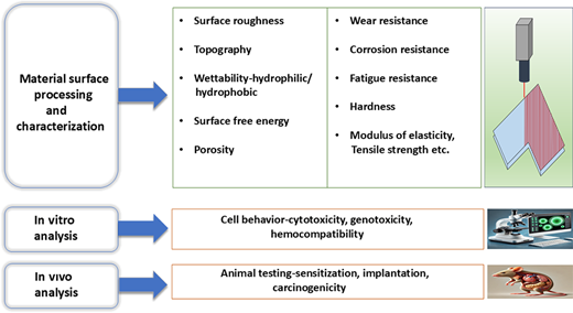
The quality of the produced geometry is impacted by factors including laser intensity, frequency, pulse duration, and scanning velocity. Refining laser parameters can achieve intricate geometric shapes with high accuracy, swift material elimination, and a confined heat-affected area.58 Iaroslav et al.59 demonstrated that laser-induced surface modifications on titanium alloys significantly improve biocompatibility by altering the surface topography. These modifications enhance cellular responses and tissue integration, making LST a valuable tool for biomedical applications. A diverse range of laser technologies, including excimer, fibre, Nd:YAG, CO2 lasers, Ti:sapphire, and Yb:YAG, are utilised in the laser surface treatment of biomaterials. The processes listed in Figure 7 are involved in biomaterial surface texturing research.
Pereira et al.60 observed that the augmentation of surface wettability through laser ablation, in conjunction with thermal treatment, represents a promising means of producing hydrophilic surfaces on ceramic implants. Cunha et al.61 demonstrated that the ultrafast LST technique applied to Ti-6Al-4V elicits changes in cytoskeleton morphology, distribution, and area of focal adhesion points, as well as the proliferation of human mesenchymal stem cells. Furthermore, this method results in an augmentation of surface wettability. A recent clinical study by Gholam et al.62 demonstrated that laser-textured titanium surfaces in dental implants improved osseointegration by 60%, significantly reducing implant failure rates. The laser-textured surfaces promoted faster tissue integration, enhancing the long-term success of the implants in clinical applications. Deepak et al.63 employed nanosecond laser texturing to create a pit structure for a superhydrophobic titanium alloy grade 5 Ti-6Al-4V as an antibacterial surface followed by annealing. Xinhui et al.64 highlighted the antibacterial potential of femtosecond laser-induced surface patterns. Their study showed a 75% reduction in bacterial colonisation on laser-textured orthopaedic implants, demonstrating LST’s effectiveness in preventing post-surgical infections. These findings provide promising applications for reducing bacterial growth and improving implant safety.
Stango et al.65 applied hydroxyapatite (HAp) coating to implants made of 316LSS and Ti-6Al-4V, revealing that the laser-textured surface offers superior corrosion resistance and is also suitable for biomedical purposes. Chen et al.66 utilised laser technology to produce a microhexagonal array on Ti-6Al-4V samples inspired by the adhesive skin structures observed on tree frog toe pads. The resultant surface topology comprises hexagonal cells distinctly separated by deep channels, augmenting osteoconduction properties. Prominent biomaterials frequently utilised, including titanium and its alloys, cobalt–chromium alloys, stainless steel, aluminium oxide and zirconium oxide ceramics, and ultra-high-molecular-weight polyethylene (UHMWPE), are scrutinised in terms of augmenting surface attributes.
7. LST for biomedical applications of titanium and its alloys
Since the emergence of the 1970s, materials such as titanium (including Ti alloys) have been widely utilised in biomedical applications and are particularly appropriate for dental implantology due to their exceptional specific strength, minimal electrical conductivity, and resistance to corrosion.67,68 Due to their superior specific strength, these are favoured for implants in the hip, knee, wrist, shoulder, and spine. As titanium possesses an elasticity modulus that closely approximates natural bone, stress shielding is mitigated.69 The surface profile’s topography of the implant plays a crucial role in determining the type and extent of interaction between titanium and host tissue.70 Titanium alloys are utilised in diverse biomedical domains, including bone plating, prosthetic hip and knee joint replacement, fracture treatment screws, pacemaker implants, cardiac valve prosthesis, and even the manufacture of artificial hearts.71 These alloys are highly favoured in various medical applications, including orthopaedic wire leads, cardiovascular and vascular stents, and components of heart valves.72 Due to the fascinating attribute of pure titanium, it promptly generates a slender passive oxide layer (2–10 nm) upon exposure to air in approximately 9–10 s. This protective layer shields the material from chemical erosion and corrosion, rendering it highly compatible with biological systems.18,73,74 The metal ablation procedure results in the microstructure refinement, leading to a substantial enhancement in both hardness and surface Young’s modulus.70 Although the material boasts exceptional thermo-physical properties and high biocompatibility due to its superior friction coefficient and low wear resistance, its range of applications is unfortunately restricted. The material’s surface treatment plays a vital role in addressing this issue. Researchers have employed various methodologies globally to treat the surfaces of titanium and its alloys.75 Ti material (grade 2) was subjected to shot peening, acid etching, and laser treatment. This demonstrated the shift in surface chemistry from hydrophobic to hydrophilic, and most protein adsorption was seen in the laser-textured surface.76 Laura et al.54 fabricated different cross-hatched micropatterns on Ti-6Al-4V, studied wettability and friction against bone, and found that distinct patterns influence these factors. Bahera et al.77 noted the existence of biphasic calcium phosphate deposition in textured Ti-6Al-4V materials, which led to increased bioactivity, proliferation, and cell adhesion that could be beneficial for dental and orthopaedic uses. Wang et al.78 conducted a study investigating the effect of microgroove width on both anti-corrosion and bio-tribological properties of Ti-6Al-4V-graphene oxide coating. The results showed significant improvement in both anti-corrosion and anti-wear characteristics. Figure 8 presents a schematic illustration of the solid ultraviolet laser with a maximum power output of 5 W, which operates at a wavelength of 355 nm. A laser is produced at the laser head, followed by two galvanometric mirrors directing the resulting beam. Subsequently, an F-theta scanning lens with a sophisticated engineering design effectively transfers the laser beam onto the workpiece material surface for optimal efficiency during the laser texturing process. The focusing conditions are determined by measuring the distance between the F-theta lens and the workpiece material surface. It was reported that for Ti-6Al-4V, increasing groove width and pitch can enhance surface wettability. The superimposed framework fabricated on the Ti alloy enhances its mechanical properties while simultaneously refining the grain structure of the alloy. Zheng et al.79 have reported that increasing the width and pitch of grooves can improve the surface wettability for Ti-6Al-4V. Virginie et al.80 fabricated a hybrid topography consisting of micropatterns and nanoripples using femtosecond laser technology, which was found to enhance the osteogenic differentiation of mesenchymal stem cells while suppressing their adipogenic potential. Deepak et al.63 rapidly produced a pit structure on grade 5 Ti-6Al-4V titanium through nanosecond pulsed technology, followed by annealing to create a superhydrophobic surface with antibacterial properties. Figure 9 shows SEM pictures of plain and textured titanium with various-sized pits caused by lasers. Unrefined Ti-6Al-4V and treated Ti-6Al-4V surfaces, featuring pits that are separated by distances of 40, 60, and 80 µm, respectively, are depicted in Figure 9(a). In addition, magnified images of the textures can be found in Figures 9(b) and 9(d). Hierarchical structure is the term used to describe these groupings of structures. Due to the 40 µm sample’s smallest pit spacing compared with other samples, such hierarchical structures are more apparent.
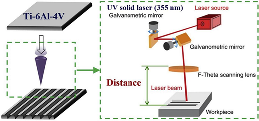
A diagrammatic illustration of the laser treatment applied to Ti-6Al-4V for the purpose of creating a microgroove is presented. This figure has been reproduced from Wang et al.,78 with permission obtained from Elsevier in 2020
A diagrammatic illustration of the laser treatment applied to Ti-6Al-4V for the purpose of creating a microgroove is presented. This figure has been reproduced from Wang et al.,78 with permission obtained from Elsevier in 2020
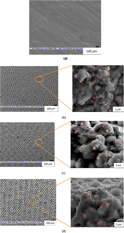
SEM images illustrating (a) the unmodified surface of Ti-6Al-4V, and (b)–(d) pit structures generated by laser with different inter-spacing distances of 40 μm, 60 μm, and 80 μm on Ti-6Al-4V.63
SEM images illustrating (a) the unmodified surface of Ti-6Al-4V, and (b)–(d) pit structures generated by laser with different inter-spacing distances of 40 μm, 60 μm, and 80 μm on Ti-6Al-4V.63
The researchers have produced bioactive Ca-P coatings that are textured and possess thicknesses of 100 and 200 m to be applied on Ti-6Al-4V substrates. The authors exhibited an augmented cytoskeleton connection and proliferation in mouse MC3T3-E1 cells resembling osteoblasts. Moreover, their investigation augmented in vitro bioactivity and biocompatibility.75 Cellular cultivation suggests that LST on Ti alloy produces microgrooves, which significantly promote the proliferation and differentiation of MC3T3-E1 cells, indicating an increase in bioactivity.81 Zhou et al.82 revealed that the utilisation of a 10-picosecond infrared laser to texture TI-6Al-4V alloy surfaces with groove arrays measuring 30 and 60 μm in depth led to substantial enhancements in both cell proliferation and adhesion. The titanium alloy underwent surface modification by way of Nd:YAG laser treatment, revealing that the smooth texture may facilitate more excellent cell adhesion due to heightened surface tension and lowered contact angle.83 Moreover, laser thermal spraying at 140 J-cm2 significantly improved the bioactivity and wettability of the titanium alloy surface compared to the unprocessed sample. Ti-6Al-4V alloy is appropriately modified by the Nd:YAG laser’s optimum parameter. The histopathological assessment, SEM analysis, and contact angle measurement collectively validate the feasibility of producing a titanium alloy with enhanced chemical and physical characteristics for deployment in biomedical settings.84 Jeong et al.85 conducted research on femtosecond laser texturing to create a nanostructured thin film on Ti35Nb-xZr Ti alloy. This resulted in noteworthy cell proliferation and spreading and an inclination towards surface wetting. The implications of these findings indicate that ternary Ti alloys displaying nanotubular surfaces possess immense potential as implant materials.
Chen et al.66 have ingeniously crafted an assemblage of microhexagonal lasers that emulate the surface architecture of hexagonal cells, demarcated by profound channels on Ti-6Al-4V samples. The objective is to augment osseointegration and osteoconduction by taking cues from the adhesive skin structures observed in tree frog toe pads. Figures 10(a–d) exhibit the surface microhexagonal configurations on Ti-6Al-4V samples, featuring hexagons with side lengths of 150, 200, 250, and 300 m. The depiction reveals that these microhexagons exhibit coarser surfaces along their edges. Ti-6Al-4V samples exhibiting microhexagonal architectures underwent a chemical surface modification process, which included acid etching and alkaline-heat treatment, to produce the hierarchical micro/nanostructures. Figures 10(e–h) depict the surface topography of Ti-6Al-4V samples featuring microhexagonal structures that underwent chemical surface treatment, with hexagon side lengths varying from 150 to 300 m. The analysis reveals the intricate and delicate refinement of laser parameters that can result within the microhexagons. Figures 10(i–j) present a high-power microscopic view of the hexagon’s surface layer, showcasing an array of sub-microscale protuberances and holes. In contrast, Figure 10(k) features several microscale protuberances. Furthermore, Figure 10(l) presents a tri-dimensional illustration of the hierarchical surface topography observed in Figure 2(f), unveiling that channels exhibit an average depth of approximately 10 m. In the interest of clarity, we shall designate samples consisting exclusively of microhexagons produced through laser texturing as ‘microhexagons’. In contrast, those exhibiting hierarchical structures derived from a fusion of laser texturing and chemical treatment will be denoted as having ‘hierarchical structures’.
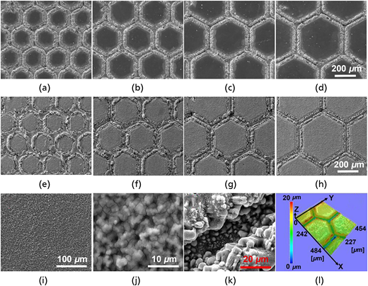
(a–d) SEM images of surface microhexagon structures on Ti6Al4V samples at different side length of hexagon: 150 μm, 200 μm, 250 μm, and 300 μm, respectively, fabricated by laser pulses at same influence of 2000 J/cm2 in the moving speed of 100 mm/min (Scale bar is 200 μm in the subfigures); (e–h) SEM images of surface morphology of Ti-6Al-4V samples with microhexagon structures after chemistry surface treatment at different side length of hexagon: 150 μm, 200 μm, 250 μm, and 300 μm, respectively (scale bar is 200 μm in the subfigures); (i) SEM image of surface morphology within microhexagon in (f); (j) SEM image of surface morphology in the high power microscopic view of (i); (k) SEM image of surface morphology in the channel between microhexagons in (f); (l) three-dimensional morphology of (f).66
(a–d) SEM images of surface microhexagon structures on Ti6Al4V samples at different side length of hexagon: 150 μm, 200 μm, 250 μm, and 300 μm, respectively, fabricated by laser pulses at same influence of 2000 J/cm2 in the moving speed of 100 mm/min (Scale bar is 200 μm in the subfigures); (e–h) SEM images of surface morphology of Ti-6Al-4V samples with microhexagon structures after chemistry surface treatment at different side length of hexagon: 150 μm, 200 μm, 250 μm, and 300 μm, respectively (scale bar is 200 μm in the subfigures); (i) SEM image of surface morphology within microhexagon in (f); (j) SEM image of surface morphology in the high power microscopic view of (i); (k) SEM image of surface morphology in the channel between microhexagons in (f); (l) three-dimensional morphology of (f).66
8. Zirconia (ZrO2) as a biomaterial
Zirconia (ZrO2) is a ceramic biomaterial increasingly substituting metal in the biomedical field for various applications, including knee and hip implants and prosthesis constructions.86–88 The complete stabilisation of zirconia chemical composition has been demonstrated by incorporating 2–3 mol% yttria (Y2O3), producing tiny 100% meta-stable tetragonal grains.89–91 Due to their osseointegration capacity, zirconia implants have emerged as a titanium implant substitute. The 3Y-TZP, a tetragonal zirconia polycrystal stabilised with yttria at 3%, has garnered recognition for its exceptional mechanical attributes, particularly its impressive flexural strength that can reach up to 900–1200 MPa,92 superior resistance to fracture with toughness ranging from 7–10 MPa/m2, outstanding ability to resist wear and tear, remarkable optical and biological properties, high level of biocompatibility, and reduced bacterial affinity.89,92–95 Moura et al.96 presented their research on laser surface treatment as applied to zirconia implants, demonstrating that the textured surface enhances chemical bonding between bone and 3Y-TZP disks. This method effectively fosters optimal bone adherence onto the implant’s surface. Faria et al.97 have devised an innovative technique for functionalising implant surfaces, which involves producing HAp-coated zirconia structures through a hybrid laser process. The microstructure was generated with an Nd:YAG laser, while the CO2 laser was responsible for sintering the HAp coating onto the surface. This method yielded structured surfaces rich in HAp and effectively prevented detachment from the zirconia surface following implantation, promoting successful osseointegration. These positive results suggest that this proposed surface design could enhance the bioactivity necessary for successful osseointegration of zirconia-based implants by solving the issue of coating separation during implant insertion. The subsequent procedures were employed to fabricate the structured samples of zirconia coated with HAp: frigid compression, laser texturisation of surface, customary sintering, immersion coating with HAp, and ultimate laser sintering to coat HAp. The various methodologies utilised in producing HAp-coated zirconia structured samples are depicted in Figure 11. After subjecting these samples to friction testing against bone submerged in PBS and an exerted force of 100 N, Figure 12 presents exceptional SEM micrographs of HAp-coated zirconia structured surfaces. These images are accompanied by corresponding EDS spectra about designated zones (Z1 and Z2). The transference of matter from the osseous tissue, depicted by the shaded regions, to the zirconia-structured surfaces coated with HAp, is discernible in the comprehensive depiction presented in Figure 12(a). Consequently, adhesion was the predominant mechanism between the contacting materials during friction testing, driven primarily by chemical affinity. Madeira and colleagues98 fabricated zirconia nanoparticles (Aunps and Agµps) that were functionalised using advanced techniques such as spray deposition, laser adhesion, texturing, and additive laser methods. These materials exhibit excellent performance in friction testing, demonstrating that their integrity remains unaltered upon implant attachment. Implementing Nd:YAG laser facilitated the creation of surface texture, ultimately boosting the mechanical interlocking between Hap powder and zirconia and subsequently intensifying its adhesion.99
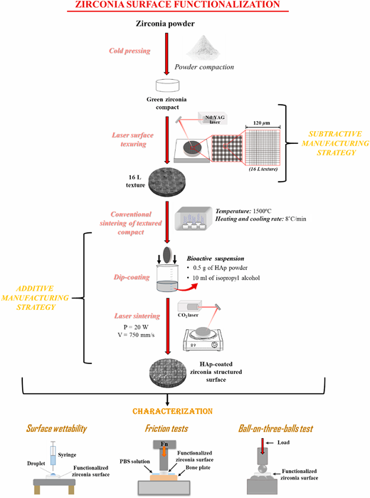
Illustration showcasing the methodology and analytical progression of zirconia structured surfaces coated with HAp.97
Illustration showcasing the methodology and analytical progression of zirconia structured surfaces coated with HAp.97
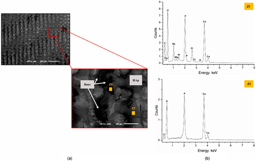
SEM micrographs of zirconia surfaces coated with HAp, following friction tests against bone (a), and the corresponding EDS spectra for labelled zones Z1 and Z2 (b).97
SEM micrographs of zirconia surfaces coated with HAp, following friction tests against bone (a), and the corresponding EDS spectra for labelled zones Z1 and Z2 (b).97
Delgado-Ruiz et al.100 conducted a study to assess the suitability of utilising femtosecond laser technology for microtexturing the surface of cylindrical zirconia dental implants. The study involved 66 zirconia implants, divided into three groups based on their surface characteristics: untreated, microgrooved textured, and those exhibiting both features. The results demonstrated that femtosecond laser microtexturing is an attractive alternative to traditional treatments due to its superior precision and minimal impact on surrounding tissues.101 Figure 13 displays the SEM images of well-designed, reproducible and equally spaced textures of zirconia at various laser powers, strategies, number of passages and scan speeds. A research study assessed the effectiveness of femtosecond laser ablation in producing micro- and nanoscale structures that can alter the surface topography of alumina-toughened zirconia. The treated surface exhibited a significant increase in the expression of osteogenic transcription factors and genes, forming a mineralised extracellular matrix compared with its untreated counterpart.
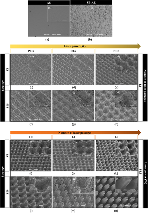
The SEM micrographs illustrate the zirconia surface, specifically (a) and (a1) as well as (b) and (b1) SB-AE. Laser-generated textures obtained through strategy Z8 or Z16 exhibit variations based on laser power, which is indicated by images (c–h) and (c1–h1).101 The number of laser passages remains constant at L1 in these images. Furthermore, the impact of varying numbers of laser passages while maintaining a constant laser power of P1.5 is explored through images (i–n) and (i1–n1).101
The SEM micrographs illustrate the zirconia surface, specifically (a) and (a1) as well as (b) and (b1) SB-AE. Laser-generated textures obtained through strategy Z8 or Z16 exhibit variations based on laser power, which is indicated by images (c–h) and (c1–h1).101 The number of laser passages remains constant at L1 in these images. Furthermore, the impact of varying numbers of laser passages while maintaining a constant laser power of P1.5 is explored through images (i–n) and (i1–n1).101
9. Laser texturing of polymers
Polymers are natural or synthetic compounds consisting of enormous molecules resulting from merging numerous monomers. These multi-faceted substances possess extensive utility in the realm of biomedical applications, with polyetheretherketone, UHMWPE, polypropylene, acrylic bone cement, and nylon being among the prevalent polymers employed for medicinal objectives.102,103 The application of UHMWPE in total hip replacement (THR) and total knee replacement (TKR) has been unequivocally established. UHMWPE is a biocompatible material that exhibits remarkable resistance to abrasion. Riveiro et al.104 propose that employing k = 532 355 nm is more advantageous in augmenting the UHMWPE polymer’s surface roughness and wettability. The CO2 laser texturing of poly (L-lactide) surfaces results in significant modifications to the mechanical properties of the treated polymer surface while concurrently adjusting its surface’s physical and structural attributes to conform with cellular specifications.105
Koufaki et al.106 conducted a study investigating the adhesion and viability of cells on polymeric surfaces featuring high roughness, gradient roughness ratio, and wettability achieved through laser micro/nanotextured Si surfaces. The findings revealed that both types of cells exhibited superior adhesion to the microstructured surfaces compared with the unstructured surface. In addition, PC12 cells demonstrated strong adherence to the patterned surface. Okoshi and Inoue107 conducted a study using femtosecond laser sources to ablate and modify the surface of polyethylene samples – the investigation aimed to examine the effect of laser wavelength on surface topography and chemistry. LST was carried out using a femtosecond Ti:sapphire laser source at 790 nm and second harmonics at 395 nm. Both wavelengths demonstrated successful ablation of polyethylene surfaces, with an ablation threshold of 50 mJ/cm2 for 790 nm and 17 mJ/cm2 for 395 nm.
Fayou et al.108 utilised a q-switched Nd:YAG laser to generate periodic lines and micropatterns on polycarbonate, employing laser interference lithography for expediency. Furthermore, high-performance fibers were analysed to demonstrate their parallel growth concerning the line structure and their bipolar morphology about point micropatterns. Aguilar et al.109 employed a femtosecond laser to generate micropatterning of polycaprolactone and polyglycolic acid in an ambient atmosphere. They fabricated microchannels and perforations that were quantified in micrometres within biodegradable microdevices. Moreover, they etched microfeatures into the polymers with a size of 30 μm. Duncan et al.110 fabricated microgrooves with dimensions of 30 by 10 μm, employing a power density of 1 J/cm2 at a wavelength of 248 nm and pulse duration of 20 ns. Their findings revealed that osteoprogenitor cells exhibit adhesion on the surface of polyethene terephthalate (PET) when grooves are minimally wide and deep. Nevertheless, the bottom portion of these grooves experienced limited cell adherence due to surface chemistry effects.
10. Laser texturing of other materials
Stainless steel is renowned for its biocompatibility, exceptional hardness, superior manufacturing capacity, and cost-effectiveness as an implant material. AISI 316L is the favoured option for structural applications and in fabricating transitory medical apparatuses such as screws, plates, hip nails, and THR uses.57,111 Lazzini et al.112 developed the model to enhance comprehension of the interplay between coccoid bacteria and surfaces exhibiting nanoripple textures. Magnesium (Mg) is biodegradable when used as an implant. As a result, it avoids the need for additional surgery to remove implant material. Furthermore, urine is used to extract biodegradable Mg. Consequently, Mg alloys, including bone plates and pins, are utilised as transitory orthopaedic apparatuses. Furthermore, owing to their elastic modulus being in the closest vicinity to that of genuine bone, they assuage any apprehension regarding stress shielding.113 Hu and colleagues.114 employed laser modification on the Mg-6Gd-0.6Ca alloy to investigate cellular responses. Their study revealed that MC3T3-E1 cells displayed enhanced adhesion to the laser-treated surface, indicating its potential as a promising technique for improving biocompatibility in biomaterials. Ma and colleagues115 have discovered a Mg-Gd-Ca surface that exhibits reduced susceptibility to corrosion following laser treatment due to modifications in the solidification microstructure.
Implementing cobalt–chromium alloys proves to be immensely advantageous in applications like THR cups and femoral heads due to their exceptional attributes such as formidable wear resistance and fatigue strength.116 Qin et al.117 employed LST to generate diverse textures on Co-Cr-Mo alloy surfaces. Their research demonstrated that the textured surfaces of Co-Cr-Mo alloys significantly impact osteoblast proliferation, gene expression, and topography. Qin et al.118 created a variety of wetting surfaces on Co-Cr-Mo using LST in combination with fluoroalkyl silane treatment. The relationship between these wetting surfaces and their tribological properties has been thoroughly examined.
A detailed comparison of different surface texturing techniques, their respective advantages, and research findings is presented in Table 2, which provides an overview of the latest advancements and key contributions in this field.
Surface texturing of biomedical implants: research overview
| Researcher/group | Focus | Year | Key findings |
|---|---|---|---|
| Eisenbarth et al.119 | Sandblasting and acid etching of titanium with different grit sizes. | 2004 | Controlled surface roughness influenced cell adhesion and differentiation |
| Hongjian et al.120 | Electrophoretic deposition of hydroxyapatite on titanium for enhanced bone bonding | 2011 | Bioactive coating improved implant fixation and osseointegration |
| Vanithakumari et al.121 | Acid etching and anodisation of titanium for micro/nanoroughness | 2013 | Hierarchical surface topography promoted osteoblast adhesion and differentiation |
| Alex et al.122 | Plasma electrolytic oxidation of titanium for enhanced bioactivity and bone integration | 2014 | Increased surface area and roughness promoted osteoblast adhesion and differentiation |
| Cunha et al.123 | Femtosecond laser texturing of titanium for enhanced osteoblast adhesion and differentiation | 2015 | Optimised surface roughness and wettability improved cell response and bone-to-implant contact |
| Zhou et al. 82 | Investigating the effect of laser texturing parameters on the biocompatibility of titanium implants | 2017 | A comprehensive study on parameter optimisation for optimal cell-material interactions |
| Aliya et al.124 | Sandblasting and acid etching of stainless steel for improved corrosion resistance | 2018 | Optimised parameters led to increased implant longevity and reduced inflammatory response |
| Elisabetta et al.125 | Sol-gel coating of titanium with bioactive glass for improved osseointegration | 2020 | Surface modifications promoted tissue integration and improved implant performance |
| Zhou et al. 47 | The effect of picosecond laser texturing on titanium for enhanced wear resistance and corrosion protection | 2020 | Enhanced mechanical properties extended implant lifetime and improved biocompatibility |
| Ahmed et al.126 | Nanosecond laser texturing of Ti-6Al-4V for antibacterial properties | 2020 | Specific micropatterns inhibited bacterial adhesion and biofilm formation |
| Bonse et al.127 | Picosecond laser-induced periodic surface structures (LIPSS) on titanium for controlled cell alignment | 2021 | LIPSS orientation influenced cell morphology and migration, potentially improving implant integration |
| Wang et al.128 | 3D printing of porous titanium scaffolds with controlled architecture | 2022 | Customised scaffold design promoted bone ingrowth and vascularisation |
| Julio et al.129 | Laser surface modification of zirconia for improved wear resistance and osseointegration | 2023 | Hybrid micro/nanotextures enhanced implant longevity and bone response |
| Maria et al.130 | Chemical etching of titanium for creating antibacterial surfaces | 2024 | Specific micropatterns reduced bacterial colonisation and biofilm formation |
The evolution of LST techniques and their increasing adoption in biomedical applications are illustrated in Figure 14, showcasing the research trends from 2011 to 2024.
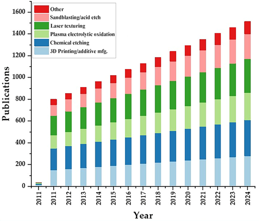
Trends in surface texturing of biomedical implants (2011–2024). Data sourced from systematic literature searches in Web of Science and Scopus, using keywords including surface texturing by chemical etching, plasma electrolytic oxidation, 3D printing/additive manufacturing, laser texturing, and sandblasting/acid etching
Trends in surface texturing of biomedical implants (2011–2024). Data sourced from systematic literature searches in Web of Science and Scopus, using keywords including surface texturing by chemical etching, plasma electrolytic oxidation, 3D printing/additive manufacturing, laser texturing, and sandblasting/acid etching
11. Conclusion
This extensive review paper thoroughly examines the applications, methodologies, and innovations in laser texturing as applied to biomedical engineering. The paper begins by elucidating the fundamental principles of laser texturing, delving into the diverse types of lasers and their interactions with different materials, including metals, polymers, and ceramics. The review’s primary focus is on the manifold applications of laser texturing in the biomedical sector. It explores how laser texturing has been employed to enhance biocompatibility, promote cell adhesion, and modulate the mechanical properties of biomedical implants. The writers deliberate on the crucial function of surface topography in dictating cellular activities, encompassing adhesion, proliferation, and differentiation. They further explore how laser texturing techniques can meticulously engineer these surface attributes to enhance biological responses. Furthermore, extensive research and development efforts have investigated various materials suitable for biomedical applications. These investigations have emphasised the significant advancements in life science technology.
Despite the copious research conducted on the application of titanium and its alloys, as well as zirconia, in biomedical contexts, there has been a dearth of examination into the effects of laser parameters on the process of laser texturing. A comprehensive analysis of diverse surface properties – roughness, wettability, and hardness – through parametric assessment is crucial for enhancing overall processes and expanding commercial applications in this burgeoning field.
Further investigation is required to delve into the physics of LST concerning other materials, and clinical trials must be conducted to validate its practicality for implementation within the biomedical field. In addition, LST has been recognised as a promising method for the alteration of biomaterial surfaces and can be efficiently utilised in biomedical applications.
12. Limitations and future scope
12.1 Limitations
While LST has shown promising results in enhancing biocompatibility, antibacterial properties, and osseointegration, several limitations still exist. One of the primary challenges is the scalability of LST for mass production in clinical settings, especially when dealing with complex geometries or large implants. In addition, the long-term clinical performance of LST-treated implants remains underexplored, as most current research focuses on short-term in vitro or in vivo studies. The variability in outcomes based on different laser parameters and material types also presents a challenge, as the ideal settings for one material may not be optimal for another. Furthermore, standardisation of laser parameters across different research studies is lacking, making it difficult to compare results.
12.2 Future scope
Future research should aim to optimise laser parameters for different biomaterials, ensuring that LST can be applied across a wide range of medical implants. Large-scale clinical trials are necessary to validate the long-term benefits of LST, particularly in reducing implant failure rates and preventing bacterial infections. In addition, integrating LST with emerging technologies, such as intelligent implants or nanotechnology, could further enhance the functionality of biomedical devices. Exploring LST’s cost-effectiveness and commercial scalability will also be crucial for its widespread adoption in clinical practice.


